Choose timezone
Your profile timezone:
The next Coherence conference in 2026 will be hosted in Wisconsin and co-chaired by Stephan Hruszkewycz (ANL) and Paul Evans (UW).
Here is the conference website : https://go.wisc.edu/coherence2026

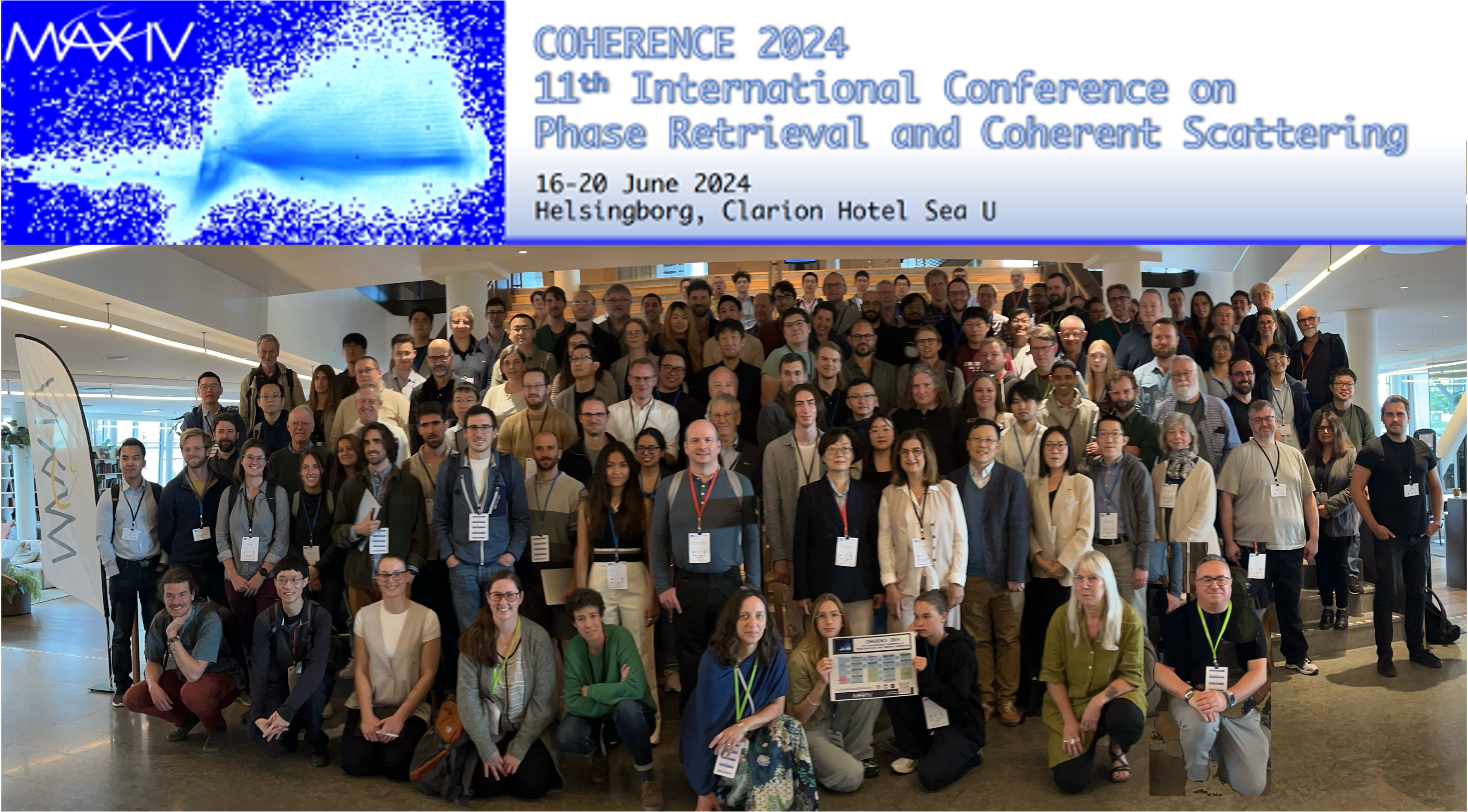
MAX IV Laboratory is pleased to host Coherence 2024 as an in-person meeting in the coastal city of Helsingborg.
The conference takes place just before Midsummer, one of the most celebrated holiday in Sweden. Book your accommodation in due time!
We are looking forward to 5 days of exciting talks and lively discussions on the science enabled by coherent methods.
The recipients of the Coherence 2024 Conferences Poster Prize are:
Congratulations to both of you !

|
|


![]()
![]()


A solid loaded beyond the yield stress loses its elastic properties and becomes plastic. From a microscopic point of view, this limit corresponds to the condition where plastic regions become so densely packed that they give rise to system-spanning structures. This limit for glasses is abrupt, which makes experimental in-vestigations challenging. Here, the yield point is reached by the alternative ap-proach of increasing the density of plastic regions by generation of point defects during x-ray irradiation. For the case of a LiBO2 glass, we show that at low doses, i.e., for a low density of defects, the defects behave as isolated stress sources that induce atomic displacements typical of an elastic solid. As the density of defects in-creases, the mechanical response of the glass at the local scale changes from elastic to more and more plastic, until reaching the limit where it becomes charac-teristic of a flowing system, which signals that the yield point is reached.
X-ray Photon Correlation Spectroscopy (XPCS) is a well-established technique to study slow dynamics in disordered materials at nanometer down to angstrom length scales [1]. The method exploits the coherent fraction of the synchrotron radiation and benefits enormously from the recent upgrade of the ESRF source (EBS) [2]. The high degree of coherence opens new avenues for application of XPCS. One of the promising avenues is the study of dynamics in concentrated protein solutions at nearest neighbor distances. Diffusion of proteins on length scales of their own diameter in highly concentrated solutions is essential for understanding biological systems such as a living cell, but its experimental characterization remains a challenge. Our work addresses this problem and discusses the use of X-ray Photon Correlation Spectroscopy at a recently upgraded 4th generation synchrotron source for this purpose. While X-ray radiation damage was generally believed to seriously
threaten the application of XPCS to biological systems, we now present a dedicated experimental and analysis strategy [3] to overcome this obstacle. We report a successful test of this approach to highly concentrated solutions of the eye lens protein alpha crystallin [4], which has previously been established as a model protein exhibiting the classic behavior of hard sphere colloids under these conditions [5]. The thus obtained intrinsic relaxation times for so-called long-time cage diffusion indeed agree with macroscopic measurements of the zero shear viscosity [6]. Our experiments also reveal a complex dependence of the key structural and dynamic properties of the protein solutions on both the total absorbed radiation dose as well as the dose rate. We discuss possible mechanisms responsible for the observed radiation effects and their consequences for future applications of XPCS.
Denaturation, aggregation and gelation of proteins and lipids are biologically relevant out-of equilibrium processes which are coupled by a hierarchy of length, time and energy scales. Finding the characteristic scaling laws governing these processes on the relevant time and length scales is necessary to predict the changes of biomolecules to future time scales.
Here, we use heated egg yolk as a model system to reveal the spatio-temporal relationships underlying these intricate processes for a wide range of time and temperature combinations [1]. Using low-dose X-ray photon correlation spectroscopy (beamline P10 at PETRA III, Hamburg, Germany) in ultra-small angle X-ray scattering geometry, we follow the time-resolved structural and dynamical evolution of multiple non-equilibrium processes occurring in a heated hen egg yolk. Following key structural and dynamical features, we identify non-equilibrium processes such as denaturation and aggregation of proteins, protein gelation, gel ageing, two-step aggregation of yolk low-density lipoproteins (LDLs), and gelation of yolk granules. We find that the overall kinetics and dynamics governing protein denaturation, aggregation, and gelation follow Arrhenius-type time-temperature superposition (TTS). This implies an identical mechanism underlying these consecutive processes, with a temperature-dependent reaction rate. At high temperatures, TTS breaks down during gelation and temperature-independent gelation dynamics is observed. This indeed reflects the complex association of protein aggregates that results in a gel network. In a broader sense, our research [1] provides an illustration of how to comprehend the fascinating non-equilibrium events in inherently complex, multi-component, thermally driven biological systems on length scales ranging from nanometers to micrometers in a time spectrum of milli-seconds to hours.
References:
[1] Anthuparambil, N.D., Girelli, A., Timmermann, S. et al. Exploring non-equilibrium processes and spatio-temporal scaling laws in heated egg yolk using coherent X-rays. Nat. Comm. 14, 5580 (2023). https://doi.org/10.1038/s41467-023-41202-z
Proteins play essential roles in life, for instance serving as carriers, participants in the immune response or for their structural role. In vivo, they exist within crowded environments with protein volume fractions typically ranging as high as 30%. When the environment becomes highly concentrated, the dynamics of proteins deviate significantly from those observed in a dilute system. However, the precise mechanisms influencing these dynamics across different time scales are not yet fully understood. Here we present our recent results [1], where we investigated the effect of self-crowding on protein diffusion in a ferritin solution with varying concentrations using X-ray Photon Correlation Spectroscopy. This technique allows simultaneous monitoring of both the structure, through small angle scattering, and the diffusion, through intensity-autocorrelation functions, of the protein solution, as demonstrated in our previous study [2]. By analyzing the scattering intensity, we observed that the ferritin particles become more densely packed with increasing protein concentration, indicated by a pronounced peak in the structure factor that shifts towards lower momentum transfer values. The protein diffusion, measured at all concentrations, follows a Brownian type of motion, but exhibits deviations at the peak position. This deviation can be attributed to the crowding effect caused by neighboring proteins, which act through hydrodynamic interactions. The hydrodynamic functions, which reflects these interactions, exhibit a peak which coincides with that of the structure factor indicating the connection of the crowding and the hydrodynamic interactions. To elucidate the underlying mechanism, we compare the hydrodynamic functions with estimations based on the δγ-theory, which considers the non-trivial interactions between particles. The model indicates that the protein diffusion is slower than that of non-interacting hard spheres due to the presence of solvent-mediated interactions and effective local friction between the particles.
[1] Girelli, Filianina et al., in preparation
[2] Reiser, Girelli et al., Nature Communications 2022,13 (1), 5528
Bragg ptychography is a phase retrieval method combining crystalline sensitivity and the possibility to image extended 3D samples [God2011, Mas2017]. Recent progresses in Bragg ptychography formalism, namely, the introduction of the back projection operator [Hru2017] and the advent of 4th generation synchrotron sources have enabled the use of 3D Bragg ptychography with unprecedented accuracy, sensitivity and spatial resolution. Specifically, the probe retrieval [Li2021] and the beam-position refinement [Li2022] are now made possible and provide improved image quality.
In this talk, I will first detail recent advance of Bragg ptychography. Using a series of preliminary results, I will further highlight the perspectives offered by 4th generation synchrotron sources, in terms of temporal resolution and strain analysis. I will finally present a project of integration of Bragg ptychography at synchrotron beamlines, performed with collaborators from ESRF, MAXIV and Diamond. It aims at enabling the use of this 3D crystalline microscopy approach to a larger x-ray community.
[God2011] P. Godard et al., Nature Commun. 2, (2011) 568
[Mas2017] F. Mastropietro et al., Nat. Materials 16, (2017)
[Hru2017] S. Hruszkewycz et al., Nat. Materials 16 (2017)
[Li2021] P. Li et al., Nat. Commun 12 (2021), 7059
[Li2022] P. Li et al., Light : Science & Applications 11 (2022), 73
Ptychography is a scanning coherent diffraction imaging technique capable of simultaneous imaging of extended samples and beam characterization with diffraction-limited resolution. It is well developed at synchrotrons, but its scanning nature prevents its use for single-shot imaging at wide applications on FEL facilities.
Single-shot ptychography can be performed by collecting the diffraction patterns of multiple overlapping beams in one shot, thus measuring the whole dataset at once and removing the need for scanning. A setup realizing this principle was proposed for visible light[1]; however, it cannot be straightforwardly applied to X-ray due to the use of refractive optics.
We solved this problem by using a single-shot ptychography setup based on a combination of X-ray focusing optics and beam-splitting grating and a corresponding forward model that facilitates single-shot imaging of extended samples at soft X-ray wavelengths [2]. The setup was tested during the proof of concept experiment at the free-electron laser FLASH at DESY and allowed us to obtain a reconstruction of a test sample and probe wavefield from the data measured with a single pulse of FLASH. However, the fidelity and resolution of the reconstruction were limited by the low performance of the diffraction grating and the inability of the forward model to fit the inter-beamlet interference.
Here, we present further progress in the single-shot psychography at FELs. We used an improved experimental setup based on the Damman grating [3] with higher diffraction efficiency and diffraction order uniformity and an improved forward model. These improvements allowed us to perform single-shot ptychographical imaging and beam characterization during the beamtime at FLASH. This technique further improved and adapted for harder X-rays, will allow the high-resolution single-shot imaging of extended dynamical samples as well as the single-shot beam characterization at X-ray free-electron lasers.
[1] Sidorenko, Pavel, and Oren Cohen. "Single-shot ptychography." Optica 3.1 (2016): 9-14.
[2] Kharitonov, Konstantin, et al. "Single-shot ptychography at a soft X-ray free-electron laser." Scientific Reports 12.1 (2022): 14430.
[3] Krackhardt, U., and N. Streibl. "Design of Dammann-gratings for array generation." Optics communications 74.1-2 (1989): 31-36.
Spin caloritronics are currently a science focus due to their potential exploitation in the next generation of spintronics applications. This class of materials combine both spintronic and thermoelectric functionalities by interconversion of charge, spin and heat currents [1]. Revealing how atomic strain and magnetic structure are intertwined at the nanoscale is of central importance to the development of emerging spin caloritronic nanotechnologies [2]. The recent investment in high brilliance 4th generation synchrotron sources hold promise for the development of new microscopic tools to reveal simultaneously atomic and magnetic nano-structure.
Here, we present preliminary results from ID01 of the ESRF-EBS and NanoMAX of MAX IV Laboratory. The first combine Bragg ptychography with X-ray resonant scattering at low temperatures to investigate prototype spin caloritronic devices structures of Gd3Fe5O12 epitaxial films capped with a Platinum layer. The second are focussed on the analysis of structure and strain of diverse prototype structures at room temperature. From our analysis exploiting inverse microscopy approaches, we demonstrate the potential to correlate atomic strain and magnetic structures down to 16 nm spatial resolution.
References
[1] S. Geprägs et al. Nature Com. 7, 10452 (2016).
[2] P.G. Evans et al. Science Advances 2020, 6 (40), eaba9351. DOI: doi:10.1126/sciadv.aba9351
M. Pancaldi, M. Fanciulli, A.-E. Stanciu, M. Guer, P. Carrara, C. Spezzani, E. Pedersoli, M. Luttmann, M. Vimal, D. Bresteau, D. De Angelis, P. R. Ribic, B. Rösner, C. David, M. Manfredda, F. Guzzi, C. B. Bevis, J. Barolak, S. Bonetti, I. Bykova, L. Novinec, A. Ravindran, A. Simoncig, D. E. Adams, G. Kourousias, G. Mancini, P. Vavassori, R. Sousa, I.-L. Prejbeanu, L. Vila, L. Buda-Prejbeanu, B. Dieny, G. De Ninno, F. Capotondi, T. Ruchon, M. Sacchi
The interaction of light beams with magnetic materials defines the rich set of analytical tools in magneto-optics, covering photon energies from infra-red to hard x-rays. In addition to the spin angular momentum (SAM) associated to the light polarization, Laguerre-Gaussian (LG) beams carry also an orbital angular momentum (OAM) of ℓℏ/photon [1] associated to an azimuthal dependence exp(iℓϕ) of the electric field phase. Over the last thirty years, OAM beams at vis-IR wavelengths found applications in biology, telecommunication, imaging and quantum technologies [2]. Their capability to exert a mechanical torque was exploited to create optical spanners for manipulating small particles. The azimuthal phase dependence introduces a singularity on the propagation axis and a radial modulation of the intensity (ring-shaped), properties that have been used to modify magnetic ordering, to improve the spatial resolution in microscopy, and to enhance the edge sharpness in phase-contrast imaging.
Over the last decade, the generation of OAM beams at shorter wavelengths, from XUV to hard x-rays, is also finding an increasing number of applications, often based on extrapolations of previous work carried out in the visible range. For instance, as it happened for the SAM, the handedness imposed by the OAM has been exploited to perform x-ray spectroscopic studies of magnetic materials [3] and of chiral molecules [4], and a recent ptychography study [5] showed that the attainable spatial resolution in the reconstructed XUV images increases with ℓ.
We will review recent extensions in the use of OAM beams from visible to short wavelengths, with focus on applications of 10-100 fs XUV-OAM pulses for element-selective spectroscopy and imaging of magnetic structures. We will show how time-resolved resonant scattering experiments offer new perspectives for tracking the dynamics of complex magnetic topologies.
Fluctuations and stochastic processes are ubiquitous in nanometer-scale systems, especially in the presence of disorder. Real-space access to fluctuating states is impeded by a fundamental dilemma between spatial and temporal resolution. Averaging over an extended period of time (or repetitions) is key for the majority of high-resolution imaging experiments, especially in weak contrast systems. If, by lack of better knowledge, averaging is indiscriminate, it leads to a loss of temporal resolution and to motion-blurred images.
We present coherent correlation imaging (CCI) [1] – a high-resolution, full-field imaging technique that realizes multi-shot, time-resolved imaging of stochastic processes. The key idea of CCI is the classification of Fourier-space coherent scattering images based on the speckle fingerprints of the real-space state – even at a low photon count where imaging is not possible. Contrast and spatial resolution emerge by averaging selectively over same-state frames. Temporal resolution down to the acquisition time of single frames arises independently from an exceptionally low misclassification rate, which we achieve by combining correlation-based similarity metric with powerful classification algorithm.
We apply CCI to study previously inaccessible magnetic fluctuations in a highly degenerate magnetic stripe domain state with nanometer-scale resolution. Our material is a Co-based chiral ferromagnetic multilayer with magnetic pinning low enough to exhibit stochastically recurring dynamics that resemble thermally-induced Barkhausen jumps near room temperature. CCI reconstructs high-resolution real-space images of all domain states by holographically aided phase retrieval [2, 3] and, unlike previous approaches, also tracks the time when these states occur. The spatiotemporal imaging reveals an intrinsic transition network between the states and unprecedented details of the magnetic pinning landscape allowing us to explain the dynamics on a microscopic level.
CCI massively expands the potential of emerging high-coherence X-ray sources and paves the way for addressing large fundamental questions such as the contribution of pinning and topology in phase transitions and the role of spin and charge order fluctuations in high-temperature superconductivity.
[1] C. Klose et. al., Coherent correlation imaging for resolving fluctuating states of matter. Nature 614, 256-261 (2023)
[2] S. Zayko et al., Ultrafast high-harmonic nanoscopy of magnetization dynamics. Nat. Commun. 12, 6337 (2021).
[3] R. Battistelli, et. al., Coherent x-ray magnetic imaging with 5 nm resolution, Optica, DOI 10.1364/OPTICA.505999
Arrays of nanoscaled magnetic elements, each acting as a single mesospin, are the building blocks of artificial systems of varying complexity in which the mesospin and lattice geometry can be used to design emergent mesoscale magnetic order. The geometry of the mesospin lattice determines the magnetic dimensionality and the interactions between the elements affect the global ordering and thermally driven dynamics [1]. Here, we focus on two types mesospin arrays arranged as Ising chains and square artificial spin ice (SASI) structures [2,3]. Different mesospin gaps generate varying interaction energies which compete with the thermally active Fe/Pd base material to drive the collective behaviour . As a function of increasing temperature, individual mesospins start to reverse, introducing defects into the arrays and reducing the correlations over characteristic timescales. Direct imaging using PEEM is limited for fluctuating systems due to long acquisition times and a limited field of view, so here we use a different approach and combine coherent magnetic scattering with x-ray photon correlation spectroscopy (XPCS). We measure one-time and two-time correlation functions as a function of applied field and temperature. We particularly concentrate on the temperature window between the mesospin blocking temperature TB (fixed by the Zeeman and shape anisotropy) and the Curie temperature of the Fe/Pd base material. This study yields new insights into the dynamics of magnetic excitations in these arrays, with both high spatial and temporal resolution.
[1] H. Stopfel et al., Phys. Rev. Materials 5, 114410 (2021).
[2] M. S. Andersson et al., Scientific Reports 6, 37097 (2016).
[3] V. Kapaklis et al., Nature Nanotech 9, 514–519 (2014).
The development of advanced functional materials relies on understanding interactions and heterogeneity at nanometer-to-micrometer length scales. The extraordinary electromechanical properties of relaxor ferroelectrics are widely attributed to the crucial role of spatial structural heterogeneity. Recent developments in coherent x-ray sources and methods significantly advance the possibilities of nanoscale measurements, offering superb spatial and temporal resolution, and support also in-situ type experimental techniques. Wide-angle X-ray photon correlation spectroscopy (XPCS) is a powerful tool to probe dynamics of heterogeneity in condensed matter, both in equilibrium and under applied stimuli [1,2]. Here, we present an in-situ XPCS study of the relaxor ferroelectric PbMg1/3Nb2/3O3 (PMN) under applied AC electric field [3]. We observed strong periodic response in two-time correlation function (TTCF) calculated from the diffuse scattering speckle pattern, even for relatively weak applied AC fields. This is surprising since PMN is electrostrictive, with no linear piezoelectric response at zero field. The periodic behavior in the TTCF was shown to arise from local tilting of the illuminated sample volume due to the combined AC field and a static field caused by the incident X-ray beam. To qualitatively describe the results (tilt amplitude and direction) we developed a model that combines the electrostrictive response of the PMN material and the non-uniform charging due to the incident micrometre-scale X-ray beam. The X-ray-induced piezoresponse may play a crucial role in interpreting XPCS and nanodiffraction studies on other insulating materials subjected to applied AC fields or varying X-ray illumination.
Work supported by the US Department of Energy (DOE), Office of Science, Basic Energy Sciences, Materials Science and Engineering Division. The experiments were performed at beamline 8-ID-E, and also 12-ID-D and 33-BM of the Advanced Photon Source, a DOE Office of Science User Facility operated by Argonne National Laboratory under Contract No. DE-AC02-06CH11357. Work used resources at the Center for Nanoscale Materials, a DOE Office of Science User Facility, undersame contract. This research was also supported by the Natural Sciences and Engineering Research Council of Canada (NSERC, Discovery Grant No. RGPIN-2023-04416).
[1] Shpyrko, O. G., et al. "Direct measurement of antiferromagnetic domain fluctuations." Nature 447.7140 (2007): 68-71.
[2] Sandy, A.R., Zhang Q., and Lurio B.L. "Hard x-ray photon correlation spectroscopy methods for materials studies." Annual Review of Materials Research 48 (2018): 167-190.
[3] Sheyfer, D., Hao Z. et al. "X-ray-induced piezoresponse during X-ray photon correlation spectroscopy of PbMg1/3Nb2/3O3." Journal of Synchrotron Radiation 31.1 (2024).
I will present recent advances in X-Ray Photon Correlation Spectroscopy (XPCS) at the MID instrument of the European XFEL. Access to sub-ms timescales is important for studying diffusion-type dynamics in aqueous solutions, for instance of colloids and bio-molecules. The European XFEL with its MHz repetition rate, provides this opportunity. XFEL experiments often require specialized sample replenishing and sample environments, for instance liquid jet delivery or fast scanning methods. The short-pulse duration and high peak brilliance of the source also gives unique possibilities for investigating the initial states of crystallization, for instance in a super-cooled liquid. X-ray Cross-Correlation Analysis (XCCA) yields direct access to studying the crystalline order and emerging defects, beyond classical crystallography experiments.
In the past decades, X-ray ptychography has been demonstrated as a powerful technique of coherent diffractive imaging because of its capability to achieve quantitative phase contrast and nanoscale resolution not limited by the performance of X-ray lenses. It has seen applications in a broad range of research fields from microelectronics to biology [1,2]. However, because ptychography is a scanning-based imaging method and requires high positioning precision of the instruments, the long measurement time and noticeable efforts needed in developing and operating the hardware have been two of the major factors limiting more general utilization of the technique. At the Hard X-ray Nanoprobe (HXN) beamline [3] of NSLS-II in the US, we have developed a system for ptychography and tomography imaging, which enables rapid scanning up to 1kHz speed and simplifies sample preparation and alignment process for more versatile applications.
The Rapid Scanning Microscopy Instrument-II (RASMI-II), being commissioned at the HXN beamline, is the main component of the imaging system which consists of motorized stages for sample and focusing optics. The sample stage uses a precision metrology disc and linear laser interferometers for position referencing, which enables continuous tracking of the sample position in 3D during scanning and sample rotation. This design largely simplifies the sample alignment procedure before measurement and tackles sample drifts during measurement to reduce scan overhead time. By combining the RASMI-II with a fast-response detector and high-speed data collection devices, we were able to push the scanning speed of ptychography measurements to 1kHz in fly-scan mode, which is the fastest frame rate of the detector, thereby achieving tomography measurement time down to 1 hour per sample. We have also developed specialized software and algorithms which incorporates fly-scan trajectories in the iterative phase retrieval process for faster convergence and noise reduction in rapid fly-scan ptychography reconstruction, as well as utilizes 3D position registration data for tomographic alignment and reconstruction.
In summary, the new ptychography and tomography imaging system at HXN beamline based on RASMI-II will provide a platform for high-throughput and versatile nanotomography imaging. Its instrument approach finds a broader potential application for other microscopy beamlines, particularly at the diffraction-limited light sources, where the scanning speed should be commensurate with significantly higher coherent flux.
[1] Holler, M., Guizar-Sicairos, M., Tsai, E.H., Dinapoli, R., Müller, E., Bunk, O., Raabe, J. and Aeppli, G., 2017. High-resolution non-destructive three-dimensional imaging of integrated circuits. Nature, 543(7645), pp.402-406.
[2] Victor, T.W., Easthon, L.M., Ge, M., O’Toole, K.H., Smith, R.J., Huang, X., Yan, H., Allen, K.N., Chu, Y.S. and Miller, L.M., 2018. X-ray fluorescence nanotomography of single bacteria with a sub-15 nm beam. Scientific reports, 8(1), p.13415.
[3] Yan, H., Huang, X., Chu, Y.S., Pattammattel, A., Nazaretski, E. and Ill, P., 2019, September. Hard x-ray nanoprobe: a scanning hard x-ray microscopy beamline offering multi-modal imaging capabilities at 10 nm. In X-Ray Nanoimaging: Instruments and Methods IV (Vol. 11112, p. 1111202). SPIE.
Soft X-ray coherent techniques have proved to be a powerful tool in the investigation of static and dynamic phenomena in solid-state quantum materials, such as ultrafast light-induced phase transitions [1], fluctuating domains [2] or memory effects [3]. One particular aspect of phase transitions is their possibility of being non-reversible and/or stochastic, which ties into mostly unexplored underlying physical mechanisms and is critical when devising functional devices that exploit the nanoscale behaviour of a given system. Coherent Diffractive Imaging (CDI) and Fourier Transform Holography (FTH) can provide the spatial resolution, contrast mechanisms and sample environments required to tackle the nanoscale repeatability of a transition under a variety of conditions. Here we show a study on the repeatability of the light induced insulator-to-metal phase transition in the prototypical material VO$_2$, showing stochastic behaviour, unstable domains with different lifetimes and permanent metallic domains stable well below the critical temperature. In addition, we will present recent advances in the coherent imaging program of the BOREAS beamline at ALBA Synchrotron, highlighting imaging on magnetic Van der Waals materials.
References:
[1] Johnson, A.S., Perez-Salinas, D., Siddiqui, K.M. et al. Ultrafast X-ray imaging of the light-induced phase transition in VO2. Nat. Phys. 19, 215–220 (2023)
[2] Klose, C., Büttner, F., Hu, W. et al. Coherent correlation imaging for resolving fluctuating states of matter. Nature 614, 256–261 (2023)
[3] Chen, X.M., Mazzoli, C., Cao, Y. et al. Charge density wave memory in a cuprate superconductor. Nat Commun 10, 1435 (2019)
Recent breakthroughs in electron storage rings have enabled a notable increase in coherent X-ray flux, promising a potential acceleration in coherent imaging speed by several orders of magnitude. However, realizing this speed enhancement requires concurrent advancements in instrumentation and computation algorithms for X-ray ptychography. At the Advanced Photon Source, we have developed high-speed ptychographic scanning that offers distinct advantages in handling decoherence effects, mitigating radiation damage, and providing capability of imaging large 3D volumes. With a prototype pixel area detector operating continuously at 20 kHz, a fast ptychographic scan was showcased at a remarkable scanning speed of 4 mm/s. Furthermore, we demonstrated high-speed ptychographic tomography on lithium nickel manganese cobalt oxide (NMC) samples with dimensions of approximately 10 microns, achieving completion within just a few minutes. These developments will unlock new possibilities for utilizing X-ray ptychography to expolre samples into the fourth dimension. This includes enabling the observation of chemical states across relevant energy edges and monitoring sample evolution in in-situ environments.
This work was performed at the Advanced Photon Source, a U.S. Department of Energy Office of Science User Facility under Contract No. DE-AC02-06CH11357.
High-resolution synchrotron experiments increasingly require advanced detection capabilities to address the challenges of high-energy coherent synchrotron radiation. Hybrid Photon Counting (HPC) X-ray detectors have emerged as pivotal tools in this context [1].The EIGER2 detector advances HPC technology with its 75 μm × 75 μm pixel size, kilohertz frame rates, negligible dead time (100 ns), and count rates up to 107 photons per pixel. Particularly for experiments aiming to exploit coherent radiation, such as Ptychography, Bragg Coherent Diffraction Imaging (BCDI) or X-Ray Photon Correlation Spectroscopy (XPCS) these advantages are crucial.
Upgrades to 4th generation synchrotron sources will significantly enhance the brilliance and coherence of X-ray beams. This increase will be most dramatic in the high-energy regime, resulting in an effective increase of coherent radiation of 2-3 orders of magnitude at X-Ray energies >20 keV or higher – depending on the synchrotron source [2,3]. Coherent scattering techniques that are so far established for X Ray energies below 12 keV will also start to become relevant at energies >20 keV which will likely enable new and groundbreaking science.
HPC detectors with high-Z sensor materials are essential for enabling these experiments. They combine (a) noise-free detection with no incoherent noise contribution, (b) fast, digital readout for fast correlation times and scanning speeds, (c) high-dynamic range to resolve both weak and strong signals simultaneously with (d) high detection quantum efficiency (DQE). Particularly for experiments carried out at >12 keV where Silicon sensors become increasingly transparent, the high DQE of high-Z sensors not only reduces the needed acquisition time, but also drastically reduces the effective X-Ray dose on the sample.
We report on the EIGER2 detectors paired with the high-Z sensor CdTe in coherent experiments, demonstrating their performance, stability and reliability. We present experimental results for both XPCS and Ptychography at >20 keV, giving a glimpse on the potential of next generation HPC detectors for future experiments utilizing high-energy coherent radiation. The presented measurements were performed at several synchrotron sources: ESRF (Grenoble, France), APS (Chicago, USA), and PETRA III (Hamburg, Germany).
References
[1] A.Förster, S.Brandstetter, and C.Schulze-Briese, "Transforming X-ray detection with hybrid photon counting
detectors”, Philos. Trans. R. Soc. Math. Phys. Eng. Sci., vol. 377, no. 20180241, April 2019.
[2] P.Willmot, et.al. “SLS 2.0 Beamline Conceptual Design Report”, 2021.
[3] “PETRA IV Conceptual Design Report”, 2023.
The recent advance of X-ray free electron laser (XFEL) opens the area of ultrafast structural dynamics with a few tens of femtoseconds time resolution due to its unique characteristics of X-rays. XFEL makes it possible to obtain critical information on the intermediate states or pathways during the phase transformation, which only measures the initial and final states with many existing techniques. In my talk, I show the results of ultrafast lattice distortions with photoexcitation of perovskite oxides by time-resolved Bragg coherent X-ray diffraction imaging by taking advantage of almost 100 % of transverse coherence available from XFEL. I will present a direct observation of initial polaron generation and relevant strain evolution in perovskite-oxide nanocrystals.
This work was supported by the National Research Foundation of Korea grant NRF- 2021R1A3B1077076.
This work proposes speckle Bragg coherent diffraction imaging (spBCDI), a new approach to improve the time-resolution in BCDI microscopy. The method uses structured (or speckled) illumination to induce a convolution of the 3D frequency content associated with the nano-sized crystal with a broad kernel, acting partially along the rocking-curve (RC) direction. Such a convolution encodes 3D structural information about the sample in each 2D image, and hence significantly reduces the required oversampling ratio along the RC direction (i.e., with respect to the Nyquist frequency).
Extensive numerical simulations demonstrate data spBCDI obtained with low oversampling ratios (i.e., < 2) along the RC yield reliable reconstruction of the crystal phase. Such reductions in the oversampling ratio reduce the measurement times which are between 1.5 – 14 times (depending on the incidence angle and the speckle size) shorter than for standard (plane wave) BCDI [1], enabling spBCDI for imaging time-evolving systems in the 0.5 - 1s time scale at 4th generation synchrotrons. The simulations also explore the limits of spBCDI by evaluating the minimum oversampling ratio along the RC which still yields an invertible data set [1]. This new method is also remarkably well suited for robust imaging of strongly distorted crystals, which remains challenging in plane wave BCDI [1].
A proof-of-concept experiment has been performed at the ID13 beamline (ESRF) [2]. The structured illumination was obtained by introducing a tailored phase-plate, that we designed to match the required speckle and beam envelope sizes (manufactured by XRnanotech GmbH, Switzerland). Combined with the powerful focusing optics of ID13 (Multi-Laue lenses), this new optical device produces a structured illumination at the sample position, with speckle grains of 70 nm laterally distributed within a beam of ~ 1 µm size. This illumination was used to image a 3D crystalline nano-structured Si thin-film with a set of oversampling ratios along the RC direction ranging from 4 down to 0.4.
[1] I. Calvo-Almazán, V. Chamard, T. Grünewald and M. Allain, Inhomogeneous probes for BCDI: Toward the imaging of dynamic and distorted crystals, in preparation
[2] I. Calvo Almazán, T, Grünewald, P. Li, P. Fenter, F. Bartolomé, M. Burghammer, A. Medjahed, M. Allain and V. Chamard, Real-time imaging of dynamic crystals with strongly structured probes for BCDI, in preparation
The study of nanoparticle (NP) assisted chemical reactions is important for the development of efficient catalyst materials for a wide range of environmental and energy applications [1]. Such studies are primarily focused on the role of surface sites - whether on particle facets or vertices. The rate and efficiency of heterogeneous catalysis is dependent on these distinct adsorption energies and turnover rates [2]. In addition to facet-dependent catalytic activity, lattice strain is known to influence the reactivity of metal surfaces [3]. One technique that can be used to probe lattice displacement with pm precision is Bragg Coherent Diffraction Imaging (BCDI) [4]. From the lattice displacement, the local strain of metal NPs can be examined in situ during catalyst-enhanced reactions. In this study, we probe the hydrogenation of acetylene on Pd nanoparticles (NP) [5], where the partial hydrogenation to ethylene is desired. This relies on fine control of the sample temperature, gas pressure, and the reactant ratios [6].
In order to probe the kinetics of heterogeneous catalysis a dynamic probe of strain evolution is found using real-time, and stroboscopic BCDI at the ID01-EBS beamline of ESRF [7]. This is applied to Pd NP in situ during acetylene adsorption, hydrogen absorption, and under acetylene hydrogenation. With this study it is possible to track the evolution of the average lattice expansion of the particle as a function of time. By distinguishing the changes in local lattice parameter under each of these conditions, we can understand the role of various Pd facets in the partial hydrogenation of alkynes [5]. With this knowledge, we performed stroboscopic BCDI on Pd NP under reversible reaction conditions to observe the mechanism with a time resolution of 0.3 s. The evolution of the lattice parameter on the sub second timescale shows a clear facet dependence which we believe is linked to the preferential adsorption of acetylene. We observe that the top (111) facet exhibits a lattice contraction relative to the bulk of the Pd NP within a few seconds upon introduction of acetylene gas. Through DFT simulations we will unravel whether the rate-limiting step is pressure stabilisation or the surface reaction with absorbed hydrogen as has been previously proposed [6].
[1] Hammer, B. & Norskov, J. K (1995). Nature 376, 238–240.
[2] McEwen, J. S. et al. (2003) Surf. Sci. 545, 47–69.
[3] Wang et al. (2016) Science 354 1031-1036.
[4] Pfeifer, M. et al. (2006) Nature 442, 63–66.
[5] Kim, S.K. et al. (2013) Jour. Catal. 306 146-154.
[6] Ravanchi, T. et al. (2018) Rev. Chem. Eng. 34(2) 215-237.
[7] Grimes, M. et al. (2024). In preparation
The recent accomplishment of a 3.88 MJ yield in 2023 along with subsequent successes at the National Ignition Facility (NIF), indicates a transformative era in Inertial Fusion Energy (IFE) research. This milestone showcases nuclear fusion’s potential as a sustainable, safe, and virtually inexhaustive source of energy, positioning it as a promising solution to meet the world’s growing energy demands. Further improvements to IFE research are dependent on assessing material response during dynamic compression. To address this, our research team is pioneering the imaging of instabilities as fusion capsules collapse. This effort aims to quantify the impact of imperfections in ablator materials during dynamic compression and how it could influence the overall fusion yield. In an experiment conducted at the Matter in Extreme Conditions (MEC) instrument at the Linac Coherent Light Source (LCLS), we employed an x-ray phase-contrast imaging (XPCI) method to accurately measure the capsule's dynamic areal density [1, 2] during this process. To mitigate the effects of random fluctuations of the x-ray free electron laser (XFEL), we implemented a flat-field correction scheme that normalizes the dynamic images, compensating for both XFEL beam inhomogeneities and imperfections accumulated along the x-ray path [3]. For quantitative evaluation, particularly for calculating the phase to determine areal density, we propose two distinct phase retrieval strategies tailored for extracting the phase from a complex, single-shot, multi-material, dynamic image. Our advanced techniques circumvent the typical limitations encountered with conventional phase retrieval methods. These traditional methods often necessitate a single-material composition, require the object to be isolated within the field of view, and are constrained to specific propagation regimes. In contrast, our approaches are not limited to these requirements and can determine the absolute phase, obviating the need for phase unwrapping. This feature is particularly beneficial since large phase excursions are induced by the compressive force of the shock wave in relation to the surrounding material. The first method involves using a single, flat-field corrected dynamic image for phase reconstruction. This method incorporates automatic differentiation (AD) [4, 5] with the transport of intensity equation (TIE), enabling the fine-tuning of specific parameters to best match experimental conditions and optimize the phase result. Further refinement of the reconstructed image is achieved by adopting the method of reconstructing the projected refractive index, a technique validated by Wittwer et al. [6] The second phase retrieval strategy leverages the inherent speckle pattern and phase contrast that naturally emerges during light propagation in the raw dynamic image. This approach avoids the conventional reliance on flat-field correction, but instead utilizes the unaltered data to uncover the phase information [7]. By using the speckle information, we reconstruct the large-scale structures, while propagation allows for the resolution of finer, small-scale features. These techniques represent a significant advancement in dynamic imaging, offering unprecedented clarity and detail in the visualization of material density. Acquiring areal density information is vital, providing insights into the complexities of achieve self-sustaining fusion reactions in current ICF experiments, has the potential to revolutionize experimental compression approaches, and can introduce additional physics to the computational models used in ICF research.
Citations:
[1] Daniel Hodge, Silvia Pandolfi, Yanwei Liu, Kenan Li, Anne Sakdinawat, Mathew Seaberg, Philip Hart, Eric Galtier, Dimitri Khaghani, Sharon Vetter, Franz-Joseph Decker, Bob Nagler, Hae J. Lee, Cindy Bolme, Kyle Ramos, Pawel M. Kozlowski, David S. Montgomery, Thomas Carver, Mathew Dayton, Leora Dresselhaus-Marais, Suzanne Ali, Richard L. Sandberg, and Arianna Gleason. Four-frame ultrafast radiography of a shocked sample at an x-ray free electron laser. In OSA Imaging and Applied Optics Congress 2021 (3D, COSI, DH, ISA, pcAOP) (2021), paper DTh7F.2, page DTh7F.2. Optica Publishing Group, July 2021
[2] K. Kurzer-Ogul, B. M. Haines, D. S. Montgomery, S. Pandolfi, J. P. Sauppe, A. F. T. Leong, D. Hodge, P. M. Kozlowski, S. Marchesini, E. Cunningham, E. Galtier, D. Khaghani, H. J. Lee, B. Nagler, R. L. Sandberg, A. E. Gleason, H. Aluie, J. K. Shang. Radiation and Heat Transport in Divergent Shock-Bubble Interactions. Physics of Plasmas, (in press), (2024)
[3] Daniel S. Hodge, Andrew F. T. Leong, Silvia Pandolfi, Kelin Kurzer-Ogul, David S. Montgomery, Hussein Aluie, Cindy Bolme, Thomas Carver, Eric Cunningham, Chandra B. Curry, Matthew Dayton, Franz-Joseph Decker, Eric Galtier, Philip Hart, Dimitri Khaghani, Hae Ja Lee, Kenan Li, Yanwei Liu, Kyle Ramos, Jessica Shang, Sharon Vetter, Bob Nagler, Richard L. Sandberg, and Arianna E. Gleason. Multi-frame, ultrafast, x-ray microscope for imaging shockwave dynamics. Optics Express, 30(21):38405–38422, October 2022. Publisher: Optica Publishing Group.
[4] Ni Chen, Congli Wang, and Wolfgang Heidrich. \partial \mathbf{H}: Differentiable Holography. Laser & Photonics Reviews, 17(9):2200828, 2023. eprint: https://onlinelibrary.wiley.com/doi/pdf/10.1002/lpor.202200828
[5] Ming Du, Saugat Kandel, Junjing Deng, Xiaojing Huang, Arnaud Demortiere, Tuan Tu Nguyen, Remi Tucoulou, Vincent De Andrade, Qiaoling Jin, and Chris Jacobsen. Adorym: a multi-platform generic X-ray image reconstruction framework based on automatic differentiation. Optics Express, 29(7):10000–10035, March 2021. Publisher: Optica Publishing Group.
[6] Felix Wittwer, Johannes Hagemann, Dennis Br ̈uckner, Silja Flenner, and Christian G. Schroer. Phase retrieval framework for direct reconstruction of the projected refractive index applied to ptychography and holography. Optica, 9(3):295–302, March 2022. Publisher: Optica Publishing Group.
[7] Andrew F. T. Leong, A Combined Speckle- & Propagation-based Single Shot Two-Dimensional Phase Retrieval Method. (2024)
In the exhibitors area
This talk will focus on coherence properties of femtosecond fluorescence produced by pumping the transition metal elements with X-ray free-electron laser (XFEL) pulses.
When the intensity of the incident beam is sufficiently low, the fluorescence will be incoherent light. The higher-order coherence of such incoherent light, especially intensity correlation between separated positions, provides rich information about the incident pulses, such as pulse duration and shape [1,2], as well as beam size on the target [3]. In the first half of my talk, I will discuss spatiotemporal diagnostics of XFEL pulses using fluorescence and their advantages over existing techniques while presenting the results obtained at SPring-8 Angstrom Compact free-electron LAser (SACLA) [4-6].
When the pump intensity is sufficiently strong to induce population inversion between inner-shell states (e.g., 1s and 2p states), the fluorescence photons become collective and exhibit directionality [7]. This coherent radiation, known as amplified spontaneous emission, can serve as the basis for an X-ray laser oscillator that generates fully coherent pulses by combining with cavity optics [8]. In the second half of my talk, I will talk about the coherence properties of amplified spontaneous emission. Based on the experimental results at SACLA and theoretical simulation, I will discuss the feasibility of an X-ray laser oscillator and requirement for X-ray optics.
[1] M. Yabashi, K. Tamasaku, T. Ishikawa, PRL 87, 140801(2001).
[2] I. Inoue et al., Phys. Rev. Acc. Beams 21, 080704 (2018).
[3] M. Yabashi, K. Tamasaku, T. Ishikawa, PRL 88, 244801 (2002).
[4] I. Inoue et al., J. Synchrotron Rad. 26, 2050 (2019).
[5] N. Nakamura et al., J. Synchrotron Rad. 27, 1366 (2020).
[6] I. Inoue et al., PRL 127, 163903 (2021).
[7] H. Yoneda et al., Nature 524, 446 (2015).
[8] A. Halavanau et al., PNAS 117, 15511 (2020).
Coherent X-ray diffraction imaging (CXDI) is a powerful method for visualizing the structure of an object with a high spatial resolution that exceeds the performance of the lens. CXDI is of several types, based on the optical systems and reconstruction methods. Plane-wave CXDI, in which a coherent planar beam is incident on a sample, can be used to observe isolated objects. Nonetheless, scanning CXDI, commonly known as ptychographic CXDI, is superior because it enables the observation of extended samples; however, its disadvantage is that improving its temporal resolution is challenging as it is based on multiframe data collection. Therefore, a method for reconstructing the image of an extended object using a single-frame diffraction intensity pattern must be established.
Previously, we proposed and demonstrated a practical method for single-frame CXDI in the hard X-ray regime [1, 2], in which a triangular aperture is used as a critical element in the optical system. The phase image of a selected field of view of an extended object was reconstructed from the single-frame diffraction intensity pattern based on a phase retrieval calculation. Furthermore, recently, we also proposed and demonstrated an approach to analyze particle motion in heterogeneous solutions over a wide spatiotemporal scale by combining XPCS and single-frame CXDI using triangular aperture [3]. By applying this approach to analyze of the dynamics of colloidal gold nanoparticles dispersed in aqueous polyvinyl alcohol solutions, we found that Brownian motion exists in the range of a few hundred nanometers, and two modes of motion exist in the micrometer range. In this presentation, the details of the CXDI method and the experimental results are presented.
References
[1] J. Kang et al., Opt. Express, 29, 1441–1453 (2021).
[2] S. Takazawa et al., Opt. Express, 29, 14394–14402 (2021).
[3] S. Takazawa et al., Phys. Rev. Res., 5, L042019 (2023).
X-ray ptychographic tomography is now routinely used at synchrotron facilities around the world, producing nanoscale resolutions in 3D. However, the acquisition times still lag significantly behind other imaging methods. As many synchrotrons upgrade to diffraction limited rings, the community is being presented with a dramatic increase in the coherent flux available. However, translating that increase in flux into increased scientific throughput remains a challenge.
The latest advances in high-speed ptychography at I13-1: Coherence of the Diamond Light Source have combined a novel acquisition scheme with the latest detector technologies to achieve a ptychography collection rate of over 100 kHz. This is providing sub-second projections and sub-hour 3D ptychographic tomography. We present the latest technical developments as well as their applications in the fields of battery materials and brain imaging.
We have developed and deployed CITIUS detectors for the XFEL facility SACLA and the synchrotron radiation facility SPring-8. At SACLA, we are building a 20.2 Mpixel detector system that can run at a maximum frame rate of 5 kHz. The CITIUS detectors for synchrotron radiation applications were demonstrated to operate at the maximum frame rate of 26.1 kHz at the full-image readout condition and have a detection capability of 1 Gphoton/s/pixel at 10 keV while keeping the single photon sensitivity. These high performances are achieved by the new integration-type pixels without charge amplifiers; it eliminates the major analog power dissipation source, thus enabling camera heads to be compact even at the high frame rate operation. Feasibility demonstration to coherent diffraction experiments was reported previously for Bragg CDI at ESRF [1] and ptychography at SPring-8 [2] with 280k and 840k pixels, respectively, and reviewed in a commentary [3].
In this talk, we report the development status of a new 5.04 Mpixel CITIUS detector for high-resolution ptychography. The system will have a clock trigger distributor to suppress any false beat signal arising from the frame rate comparable to the revolving frequency [3]. The stream data rate from the sensors reaches 30.6 petabytes/day. We report our development on the data acquisition system with FPGA cards in the edge servers and the SPring-8 data center for on-the-fly data reduction. We also briefly address the development status of a high-speed analog digitizer, which synchronously operates with CITIUS, for recording incident X-ray intensities/positions and stage positions.
References
1) M. Grimes, J. Appl. Cryst. 56 (2023) 1032.
2) Y. Takahashi, J. Synchrotron Rad. 30 (2023) 1.
3) J. Deng, A. Miceli and C. Jacobsen, J. Synchrotron Rad. 30 (2023) 859.
4) H. Nishino, Nucl. Instr. Meths. in Phys. Res. Sec. A1057 (2023) 168710.
In the restaurant on entrance floor
A challenging aspect of designing functional materials is to understand the impact of a wide range of characteristic spatial and temporal scales on stabilizing novel phases. Commonly used mean field approaches often provide reasonable estimates for static properties at macroscopic length scales. However, it does not provide the requisite fundamental insight into the important processes governing deviations from these averages where fluctuations play a critical role. Soft x-rays are a powerful element-specific probe to study such mesoscopic charge and spin textures. The coherence available at current and newly upgraded light sources can enhance our established tools to give significantly more detailed information of otherwise difficult to probe quantum states. We select the coherent part of the x-ray beam to produce an interference pattern known as speckle. Here I will discuss how we use that speckle at the Advanced Light Source to look inside, and better understand the transitions in collective electronic systems such as those of orbital ordering, metal-to-insulator materials and amorphous helical magnets1,2.
References:
[1] Singh, A., Hollingworth, E., Morley, S. A., et al., “Characterizing Temporal Heterogeneity by Quantifying Nanoscale Fluctuations in Amorphous Fe-Ge Magnetic Films”, Adv. Funct. Mater. 33, 2300224, 2023. https://doi.org/10.1002/ADFM.202300224
[2] Tumbleson, R., Morley, S. A., et al., “Nematicity of a Magnetic Helix”, arXiv:2404.13212
Ordering of large length-scale magnetic structures are commonly studied using techniques like electron microscopy (EM), small angle neutron scattering (SANS), and resonant elastic x-ray scattering (REXS). Using coherent X-rays to investigate these systems, we can probe magnetic textures in ways that were once exclusive to either EM or SANS: domain dynamics, inter-mixed phases, long and short range ordering – all with a flexible probe size (<10um). As such, coherent REXS is an important tool to extend our fundamental understanding of magnetic skyrmions, which is essential if they are to be a platform for quantum information systems. Particularly, Cu2OSeO3 (CSO) is an interesting skyrmion host material because it is an insulator – a requirement for low energy consumption devices. We will present new findings associated with skyrmion lattice (SkL) dynamics in CSO from experiments that push the collective behavior into different excited states. The insights gained from coherent REXS help to better understand domain motion, which aids in completing a microscopic explanation for the SkL dynamics.
High-speed XPCS using new coherent X-ray sources and fast detectors opens new avenues to explore fluctuation dynamics in fluids. We have been studying systems relevant to liquid-liquid extraction processes, where an organic solution of extractant molecules is used to separate ions from an aqueous solution by formation of nanoscale molecular complexes. The organic phase exhibits incipient phase separation, and critical fluctuations play a key role in the structure of the molecular complexes [1-5]. Here we present XPCS studies of microsecond timescale fluctuations within 5 K of the critical temperature Tc, carried out at APS beamline 8ID. With the 500 times higher coherent X-ray flux that will become available from the APS Upgrade, it should be possible to observe fluctuation dynamics much further away from Tc. This will enable exploration of the crossover from Ising to mean-field behavior, as well as the changes in dynamics expected at the Widom line (the locus of fluctuation maxima extending from the critical point into the single-phase region).
Work supported by U.S. Department of Energy (DOE), Office of Science, Office of Basic Energy Sciences, Chemical Sciences, Geosciences, and Biosciences Division, Separation Science Program, under Contract DE-AC02-06CH11357. This research used resources of beamlines 12-ID-C and 8-ID-I at the Advanced Photon Source, a DOE Office of Science User Facility operated by Argonne National Laboratory.
[1] D. Sheyfer et al., Phys. Rev. Lett. 125, 125504 (2020).
[2] M. Servis et al., J. Phys. Chem. Lett. 12, 5807-5812 (2021).
[3] D. Sheyfer et al., J. Phys. Chem. B 126, 12, 2420-2429 (2022).
[4] B. L. Bonnet et al., Phys. Chem. Chem. Phys. 25, 16389-16403 (2023).
[5] T. Rahman et al., J. Mol. Liq. 393, 123625 (2024).
Rare earth nickelates RNiO3 display an insulator to metal transition (MIT) which is accompanied by a magnetic transition, charge ordering, and a crystal structure change from orthorhombic low temperature to monoclinic high temperature state. While this system has been widely studied, the nature of the fluctuation across the transition, and the associated time- and length-scales is not known. Spontaneous fluctuations are important components for stabilizing topological magnetic structures such as skyrmions in quantum materials. However, the dynamic susceptibility of the nickelates remains relatively unexplored, and the role played by nanoscale phenomena such as domain-wall formation and motion, local strain fields and phase separation in underlying pathways of MIT is not well- understood. In our work, we focus on understanding the role of nanoscale heterogeneities and their fluctuations in rare earth nickelates by employing x-ray photon correlation spectroscopy (XPCS), a technique requiring highly coherent x-rays. Our XPCS measurements on NdNiO3 thin films, show complex evolution of fluctuations dependent upon temperature and wavevector q. We also observe unexpected non-equilibrium dynamics and suggests a new approach to understanding these materials.
Coherent diffraction imaging (CDI) and ptychography [1] have been widely used in X-ray synchrotron sources. The advantage of ptychography over traditional CDI is that it does not need prior information about the probe function and overcomes some of the other issues of CDI, such as non-unique solutions and a limited field of view.
In electron microscopy, ptychography has also attracted considerable interest due to its potential to achieve super-resolution [1] without using aberration correctors. Unlike conventional imaging modes, the image-forming optics of ptychography replaced by computational methods (like a ‘Digital Lens’) using an array of electron diffractions collected by fast detectors (Left). New generation of direct detection cameras are particularly suited to ptychographic 4D data acquisition with new modes of operation, such as electron counting and fast acquisition. In this talk, I will review the current development and capabilities of this emerging imaging technology (electron ptychography) in my group for light atomic detecting [2], low dose imaging [3], 3D reconstruction [4-5], coupling to spectroscopy [6], EM field mapping, and cryogenic EM [7,8], which can then tackle characterization challenges across the physical and life sciences, ranging from ferroic and battery materials to biological macromolecules.
References:
[1] Rodenburg, J. M. Ptychography and Related Diffractive Imaging Methods. 150, 87-184, (2008).
[2] Wang*, P., et al, Scientific Reports 7, 2857, (2017).
[3] Song, J., et al. Scientific Reports 9, 3919, (2019).
[4] Gao, S., et al. Nature Communications 8, 163, (2017).
[5] Ding, Z., et al., Nature Communications 13, 4787, (2022).
[6] Song, B., et al., Physical Review Letters 121, 146101, (2018).
[7] Zhou, L., et al., Nature Communications 11, 2773, (2020).
[8] Pei#, X., et al., Nature Communications 14, 3027, (2023).
Far-field electron ptychography is considered to be one of the most powerful phase retrieval techniques currently available for electron microscopists. Recently it was shown that ptychography is capable to surpass the Abbe resolution limit [1] and resolve specimens features as fine as the blurring due to the vibrations of the atoms [2]. A rather simple mathematical model of coherent diffraction pattern formation standing behind conventional algorithms [1,2,3,4] does not account for any motion or entangled states [3,4]. In order to overcome the resolution limit caused by the lattice vibrations we utilize the mixed-object formalism initially proposed in 2013 for x-Rays [4], but never applied to electron microscopy data. In contrast to conventional ptychography [1,2] that treats a single transmission function of a specimen, mixed-object ptychographic reconstruction considers a sequence of entangled transmission function states, each producing a coherent diffraction pattern via a multislice simulation [2,3]. The total diffraction pattern corresponding to the entangled system is formed as an incoherent sum over the states [3,4].
For the tests of mixed-object ptychography we used a 4D-STEM dataset [6] of a monolayer MoS$_2$ acquired in the Nion Hermes microscope at an accelerating voltage, convergence semi-angle, scan step and electron dose of 60 kV, 33 mrad, 0.2 Å and 5.3e6 $e^-/Å^2$, respectively.
To evaluate the resolution of the ptychographic reconstructions we propose a novel approach, akin to the Young fringe resolution test widely applied in TEM imaging [7]. We perform two ptychographic reconstructions with different initial guess for the object, e.g. uniform prior. Then the two independent results are shifted with respect to each other and the Fourier intensity of the difference is computed. The arising interference fringes indicate the range of spatial frequencies identical in the two results, allowing to conclude how deterministic a particular reconstruction is. Further, in contrast to conventional resolution tests, e.g. visibility of the Fourier peaks [1,2], the proposed approach allows to include aperiodic features that are crucial for a moving specimen.
Assuming a single illumination mode [2,4] we conducted pairs of ptychographic reconstructions in two scenarios: one involving a single-state and the other involving 10 entangled states of the transmission function. After 500 iterations of the gradient-descent [3] the achieved resolution was estimated. As a result, two independent pure-state reconstructions appear to produce identical information up to 1.9 $Å^{-1}$, while the mixed-object reconstructions contain deterministic information up to 2.3 $Å^{-1}$.
Even with a monolayer specimen that is not supposed to produce a noticeable amount of incoherent scattering, we show that considering multiple entangled states of the transmission function makes the underlying model more realistic and improves the quality of the ptychographic fit. Thus, we liberate ptychography from atomic vibrations, its last known resolution limit [2,3].
[1] Y. Jiang et al., doi: 10.1038/s41586-018-0298-5
[2] Z. Chen et al., doi: 10.1126/science.abg2533
[3] A. Gladyshev et al., doi:arXiv:2309.12017
[4] P. Thibault & A. Menzel, doi:10.1038/nature11806
[5] P. Godard et al., doi: 10.1364/OE.20.025914
[6] C. Ophus, doi:10.1017/S1431927619000497
[7] C. Kisielowski et al., doi: 10.1017/S1431927608080902
Multislice ptychography has been used to characterize thicker samples in 3D dimensions with spatial resolution beyond the diffraction limit. When applied to the diffraction patterns collected by scanning transmission electron microscopes, multislice ptychography reconstructs the object as a series of slices with a lateral resolution of 10s of pm and a depth resolution of 2-3 nm. Further improvements to this technique could allow atomic resolution imaging in three dimensions. Compared to electron tomography, multislice electron ptychography views the sample from only a single direction, avoiding the complexities of tilt series and image alignment and allowing for larger samples to be studied. However, the depth resolution is limiting. When viewing bulk crystalline samples from a major zone axis, most of the atoms overlap in projection and are difficult to distinguish individually. Therefore, in this work, we apply multislice electron ptychography to twisted bilayer 2D materials, tilted to minimize overlap, allowing us to extract 3D atomic coordinates for most of the atoms in the field of view. Compared against the known structure of the samples, these coordinates have sub-angstrom precision, though there are systematic issues with the coordinates along the beam direction. We also perform simulations to explore the range of experimental parameters that enable this level of depth resolution and precision. These findings underline the potential of multislice electron ptychography for determining atomic structure in 3D, even though challenges remain for extending this approach to thicker samples.
X-ray ptychography (NFP) is a coherent imaging technique widely used at synchrotron facilities, due to the ability to retrieve quantitative phase information of extended objects at a micrometric image resolution [1]. The advent of novel bright sources, an alternative to large-scale facilities, is paving the way for the translation of coherent X-ray imaging techniques outside synchrotrons and free electron lasers [2].
The ELI Beamlines facility of the Extreme Light Infrastructure ERIC near Prague has recently commissioned a laser-driven plasma X-ray source (PXS), based on a 20 mJ, sub-20 femtosecond, 1 kHz laser interacting with a copper tape to generate copper K-alpha emission at 8 keV with sub-ps pulses.
We present here a proof-of-concept for translating x-ray near field ptychography to laser-driven x-ray sources. In particular, we report the ELI PXS source characterization toward coherent diffraction imaging and the results of the first near field ptychographic imaging performed at a laser source. We discuss the results, limitations, perspectives and future developments.
[1] Stockmar et al., Sci Rep 3, 1927 (2013).
[2] Batey et al , Physical Review Letters, 126, 193902, (2021)
Speckle-based X-ray imaging (SBXI) [1, 2] is a technique that utilises random speckle modulations imprinted into an X-ray wavefront to retrieve multimodal sample information. Here, the term “multimodal” is used in the sense that SBXI can recover information regarding a sample’s X-ray attenuation, refraction, and diffusion information – three complementary signals. Requiring only a piece of sandpaper in the experimental set-up as a mask, SBXI is an appealing technique for use in a broad range of applications. Furthermore, the signal-retrieving algorithm we have developed—Multimodal Intrinsic Speckle-Tracking (MIST) [3]—makes SXBI even more appealing as it is computationally rapid yet still capable of retrieving high-quality images of the sample. Transverse speckle shifts and speckle blurring are associated with the recovered phase-shift and diffuse dark-field (DDF) signals, respectively. MIST analyses these speckle changes by considering local energy conservation for each speckle in the SBXI regime, wherein, the Fokker-Planck equation for paraxial X-ray imaging [4, 5] is combined with the geometric flow formalism for SBXI [6]. There are various iterations of the MIST algorithm [3, 7, 8, 9], with each increasing in generality by reducing the number of sample requirements. In all the published MIST approaches, the multimodal inverse problem is solved by linearising the associated Fokker-Planck equation and deriving analytical or least-square solutions for the multimodal signals. Within this presentation, a general overview of the currently published MIST approaches will be provided. This will cover the mathematical techniques utilised in solving the Fokker-Planck inverse problem, as well as the underlying assumptions. Retrieved signals from various samples imaged using a synchrotron SBXI technique will be shown. The closing section of the presentation will discuss ongoing research avenues.
[1] Bérujon, S. et al. Phys. Rev. Lett. 108(15), 158102 (2012)
[2] Morgan, K.S. et al. Appl. Phys. Lett. 100(12), 124102 (2012)
[3] Pavlov, K.M. et al. J. Opt. 22(12), 125604 (2020)
[4] Paganin, D.M and Morgan, K.S. Sci. Rep. 9(1), 17537 (2019)
[5] Morgan, K.S. and Paganin, D.M. Sci. Rep. 9(1), 17465 (2019)
[6] Paganin, D.M. et al. Phys. Rev. A, 98(5), 053813 (2018)
[7] Alloo, S.J. et al. J. Med. Imaging 9(3), 031502 (2022)
[8] Pavlov, K.M. et al. Phys. Rev. A, 104(5), 053505 (2021)
[9] Alloo, S.J. et al. Sci. Rep. 13(1), 5424 (2023)
X-ray ptychography stands out as a robust phase-retrieval coherent imaging technique, well-suited for investigating samples with diverse scale structures. However, its scanning nature necessitates a delicate balance between achieving high resolution and accommodating a large field-of-view (FOV), considering factors such as scanning time, stage travel range, etc. Typically, the FOV and resolution ratio is between 100 and 1,000. For instance, the current high-resolution ptychographic scanning using high-speed multi-pass scanning mode can attain resolution in the tens of nanometers within a 3$\mu m$ x 10$\mu m$ region in 38 seconds [1]. Nevertheless, scanning over a hundred-$\mu m$ FOV at such resolutions would extend the time required by a factor of 300. Within this timeframe, conducting ptychographic tomography on large-scale samples at high resolutions (<50$nm$) becomes impractical. A breakthrough addressing this compromise arises with the development of multibeam ptychography (MBP) [2], promising to transcend the current limitations of conventional ptychography and maximize photon utilization. MBP achieves this by employing multiple coded probes simultaneously using a 3D-printed lens array. The speed enhancement in MBP scales directly with the number of probes utilized. By overcoming the speed limitation, MBP offers the tantalizing prospect of conducting 3D nano-imaging on a large scale at high energy.
In our discussion, we will delve into the advancements in MBP development, including the maximum number of probes achieved and demonstrations with various sample systems. While the MBP method presents several opportunities, it also raises concerns regarding the comparisons with conventional ptychography and poses challenges that affect its performance in real-world experiments. Therefore, we will further discuss the quality of reconstruction comparison between single-beam and multi-beam ptychography, as well as how factors such as vibrations and photon statistics impact the data reconstruction quality.
[1] J.Deng, et al., Optics Express 30.15 (2022)
[2] M. Lyubomirskiy, et al., Sci. Rep. 12, 6203 (2022)
Incoherent diffractive imaging (IDI) is a novel imaging technique which uses the transient coherence of X-ray fluorescence to image the structure of the emitting atoms to nanometer resolution. By employing second-order spatial intensity correlations akin to Hanbury Brown and Twiss's stellar intensity interferometry, IDI retrieves the spatial distribution of the underlying emitters, facilitating high-resolution characterization of heterogeneous nanoparticles with element specificity. We present recent results from our single-particle IDI experiments at European XFEL, showing for the first time the feasibility of 3D imaging of nanoparticles using IDI.
Building upon the concept of IDI, we introduce Spectral Incoherent Diffractive Imaging (SIDI), a novel method for achieving dark-field imaging of nanostructures with heterogeneous oxidation states. With SIDI, shifts in photoemission profiles can be spatially resolved, enabling the independent imaging of the underlying emitter distributions contributing to each spectral line. In the X-ray domain, this approach offers unique insights beyond the conventional combination of diffraction and X-ray Emission Spectroscopy (XES). Our proposed method opens avenues for time-resolved, element specific and oxidation state-specific imaging of electron transfer in 3d-transition metal compounds or to study heterogeneous catalysts and battery materials where the nanoscale spatial distribution of elemental oxidation states are crucial for understanding function.
Knowledge of the structure of materials and biological samples at nanometer scales and over large volumes is essential to understand the mechanics behind their function. Coherent x-ray imaging is being developed to address this need. Far-field diffraction methods suffer from noise sensitivity and high dynamic range requirement of detector. Recently, with the rapid development of x-ray focusing elements, projection imaging provides an alternative solution with unique advantages over these limitations. Here, with our own manufactured multilayer Laue lenses, we demonstrated such a projection imaging modality at Petra III P11 beamline of DESY. An object placed just out of the focus forms a magnified hologram or projection image of the sample on a pixel-array detector. Magnifications of 30,000 or more can be obtained, meaning a 75 µm detector pixel maps to an image pixel of 2.5 nm. On the other hand, at an illumination numerical aperture of 0.014, a defocus distance of 100 um would give a field of view of 2.8 um, in single shot. This mode is particularly fast and efficient for phase-contrast imaging over a large field of view that can be easily “zoomed”. Signals contained in the holograms are boosted thousands of times with an improved signal-to-noise ratio, which makes it robust and easier to retrieve the object information from the measurements. For robust phase retrieval of the holograms, we stepped the sample relative to the probe, which was the method of near-field ptychography. A 4 nm half-period resolution imaging of hierarchical nanoporous gold has been achieved at the energy of 17.4 keV. Furthermore, a systematical numerical study was carried out to quantitatively illustrate the advantages of projection imaging modality over far-field diffraction method in terms of noise robustness and dose efficiency.
The development of high-brilliance X-ray sources, such as the fourth-generation diffraction-limited storage rings and X-ray free-electron lasers, have opened up new possibilities for X-ray imaging. X-ray Multi-Projection Imaging (XMPI)1 is a novel technique that exploits the unique capabilities of the high-brilliance X-ray sources and enables volumetric information using single pulses. Unlike tomographic experiments which record projections by sample rotation, XMPI is a rotation-free technique that split the beam into beamlets and record simultaneously multiple projections from different angles. As a result, XMPI can acquire 3D movies (4D) at least three orders of magnitude faster than tomographic methods. However, it is extremely challenging to reconstruct 4D from highly sparse projections acquired by XMPI. Deep learning's advancement offers a potential solution to this problem. Current deep-learning implementations for X-ray imaging, on the other hand, face two major challenges. First, they usually work in a supervised manner, which requires paired training datasets. Second, the robustness and reliance of such methods is not guaranteed.
This presentation will discuss how deep-learning approaches can potentially address three-dimensional (3D) and four-dimensional (4D) reconstructions for XMPI experiments by exploiting the large amounts of data provided by high-brilliance X-ray sources. We will focus on ONIX2 and 4D-ONIX3, two self-supervised deep-learning methods that reconstruct 3D/4D information from sparse XMPI projections and enhance their reliance by including the physics of the image formation. ONIX is a novel 3D reconstruction method that learns the self-consistency of sparsely recorded radiographs using physics-based neural networks. It can retrieve volumetric information from less than ten projections at previously impossible quality levels without requiring any prior knowledge. 4D-ONIX is based on ONIX, it extends the capability of ONIX by including time as a 4th dimension and applying adversarial training to enforce consistency between the measurement and the reconstructions.
References
[1] Villanueva-Perez, P., Pedrini, B., Mokso, R., Vagovic, P., Guzenko, V. A., Leake, S. J., ... & Stampanoni, M. (2018). Hard x-ray multi-projection imaging for single-shot approaches. Optica, 5(12), 1521-1524.
[2] Zhang, Y., Yao, Z., Ritschel, T., & Villanueva‐Perez, P. (2023). ONIX: An X‐ray deep‐learning tool for 3D reconstructions from sparse views. Applied Research, 2(4), e202300016.
[3] Zhang, Y., Yao, Z., Klöfkorn, R., Ritschel, T., & Villanueva-Perez, P. (2024). 4D-ONIX: A deep learning approach for reconstructing 3D movies from sparse X-ray projections. arXiv preprint arXiv:2401.09508.
Ptychographic X-ray computed tomography (PXCT) is a high-resolution imaging technique that is widely used for characterizing various materials. It has two variants, namely far-field and near-field, which, when combined with spectroscopic techniques, provide new possibilities for material characterization. PXCT can be used to analyze mass density, localize chemical elements, inspect microstructure, and even map the magnetic field of a sample. Although the acquisition of data has become faster and the size of possible scanned volumes has increased, the dream of time-resolved analysis or hyperspectral nanoimaging still requires further acceleration of data acquisition. This challenge can be addressed in two ways: either by improving the instrumentation and the geometry of the experimental setup or by modifying the data processing strategy to work with less data while still maintaining high-resolution and quantitative contrast.
Some beamlines, such as cSAXS, PSI, CH, and SWING at SOLEIL Synchrotron in France, have already adopted the first method. They offer experimental setups allowing very fast acquisition and with large beam sizes at the sample due to the long sample-to-detector distance. In this regard, we will introduce here the new French beamline, FAME-PIX, which has been in construction at the ESRF. This beamline will be dedicated to PXCT and spectro-ptycho, and it will employ an innovative scanning technology that enables quick sample scanning. We will showcase the applications of this technology by sharing the results we obtained at the SWING beamlines at SOLEIL.
Modifying the data analysis process is the second way to speed up the PXCT acquisition. We will be discussing an approach that uses Deep Learning networks based on MSDNET and TomoGAN to reduce the amount of data required by a factor of 4 or more without compromising the quality of the images. We ensure the accuracy of our results by implementing a robust refinement process that corrects any artifacts that may have been introduced by the Deep Learning networks. This approach can be applied to various tomographic techniques, and to facilitate the sharing of our neural networks with the community, we have created an AI tomographic hub called AIAX at the University of Grenoble Alpes in collaboration with other laboratories at Grenoble. The primary objective of AIAX is to assist the community in processing their data using our neural networks.
Holotomography is a coherent imaging technique that provides three-dimensional reconstruction of a sample’s complex refractive index by integrating holography principles with tomographic methods. This approach is optimal for the micro- and nano-tomography instruments at the newest generation of synchrotron sources. With the aim of facilitating the use of the optimal phase retrieval approaches, we present a family of novel algorithms wrapped in an efficient software implementation of X-ray holotomography reconstruction [1]. This development incorporates advanced iterative schemes for simultaneous object phase and beam illumination retrieval, facilitating high-fidelity reconstructions with enhanced accuracy. Notably, these schemes minimize the number of measurements, such as holography planes (distances) and tomography angles, required for accurate object reconstruction, thereby optimizing data acquisition efficiency. Basic holography and tomography operators are implemented using CUDA C-language functions, leveraging the computational power of GPUs for rapid processing. Iterative reconstruction schemes are written in CuPy, ensuring seamless integration into existing workflows. For interactive data exploration and analysis, the package is accessible through Jupyter Notebooks, well-suited for fast prototyping to advance the field of 3D phase contrast imaging. The efficacy of the software is demonstrated with experimental data from the Projection X-ray Microscope Instrument at the ID16A beamline of ESRF.
[1] https://github.com/nikitinvv/holotomo
Apart from being an indispensable tool in material sciences of outstanding spatiotemporal resolution that reaches towards 1 Å at 1 ps [1,2], electron microscopy is uniquely suited to study basic quantum mechanical aspects because the wave/particle duality of electrons can be seen as a window into quantum physics [3]. This contribution experimentally validates a model of electron self-interferences. They originate from coherent-inelastic scattering processes with energy loss ΔE = ħ/2/Δt at the Heisenberg limit, which marks a boundary between the classical mechanics of particle physics and the quantum mechanics of wave functions. Thereby, wave packages of finite widths are created with 0.5 radian decoherence phase that are characterized by a self-coherence length ls. A method is described that allows measuring ls and thereby self-coherence by the extension of an evanescent field at sample/vacuum interfaces. It is caused by the localization of wave functions upon pulse-like interaction of single electron waves with matter. Further, self-interferences of single matter waves rather than ensemble interferences of many electrons determine the occurrence of lattice fringes in high resolution images if energy losses do not exceed ≈ 100 eV, where the ability is lost to coherently illuminate crystal unit cells. At larger energy losses the wave packages are comparable to the size of atoms and even their constituents and are perceived as particles. The description of electron scattering in a wave picture is mandatory because decoherence determines the measured self-coherence length but does not exist in particle views, which is why a collapse of wave functions upon detection is often postulated.
[1] Kisielowski C, Specht P, Rozeveld SJ, Kang J, Fielitz AJ, D. Barton, Salazar AC, Oscar D. Dubon OD, Van Dyck, D & Yancey DF (2021). Microanalysis 27, 1420-1430.
[2] Chen FR, Van Dyck D, Kisielowski C, Hansen LP, Barton B & Helveg S (2021). Nature communications 12, 5007.
[3] Kisielowski C, Specht P, Helveg S, Chen FR, Freitag B, Jinschek JR & Van Dyck, D (2023). Nanomaterials 13, 971.
Crystallization is a well-established topic. It covers many life science fields, and its importance varies from understanding nanoparticle arrays to protein formations and drug engineering through molecular design. One of the most popular methods for direct imaging of crystal formation, defects, etc., is transmission electron microscopy (TEM), mainly used for inorganic materials since the electron dose is not as limiting as it is for the imaging of organic materials. While challenging, high-resolution electron microscopy of organic material is possible due to the recent revolutions of stable and sensitive detectors and microscopes. Real-space images of organic crystals and their aperiodic features obtained by a modern cryo-electron microscope potentially show the detailed structure. However, high-resolution details remain hidden because strong defocus conditions must be applied to produce contrast at a low electron dose. We used phase retrieval by focal series reconstruction (FSR) developed originally for inorganic samples. We have adapted the method for fast series recording of hundreds of frames within a few seconds on a direct-electron detector, all at low dose conditions (at the order of 101-102 e-/Å2 per acquisition) in cryogenic temperatures. After the reconstruction of the exit-plane wavefunction, we could eventually retrieve focus and two-fold astigmatism aberration-fixed phase images of small organic crystals with a resolution up to the information limit of the microscope. Implying further techniques, such as density functional theory (DFT), ultimately allows for solving a crystal structure with high confidence based on the image in real-space.
Electron microscopy is a powerful tool for imaging soft biological samples, providing superior resolution compared to both light and x-ray imaging techniques. Unfortunately, biological samples are weak-phase objects in the electron microscope, providing minimal inherent contrast [1, 2]. This is in part because our detectors are only sensitive to variations in electron intensity and not phase. There are different approaches to overcome these limitations, including heavy metal staining (mass-thickness contrast), defocus phase contrast, and phase plate induced phase contrast.
In this work we are developing thin film based phase plate devices, operating in Zernike and Hilbert mode respectively, to enhance phase contrast at minimal defocus. These phase plates introduce a phase shift of pi/2, or pi respectively, to the scattered beam. The phase shift produced by a thin film is governed by the mean inner potential of the film, the film thickness and the accelerating voltage of the electron microscope [3].
We utilize off-axis and in-line electron holography to characterize the phase shift induced by the thin film material of our devices [4, 5]. Employing the accelerating voltage, the known film thickness, and the measured phase shift, we can determine the mean inner potential for the material [6]. With the mean inner potential in hand we can now produce phase plate devices with the correct thickness to induce the target phase shift. With these devices we will be able to optimize the contrast from a variety of weak-phase objects yielding TEM images with both higher resolution and higher contrast.
References
1. R. Danev et al., Practical factors affecting the performance of a thin-film phase plate for transmission electron microscopy, 2009, Ultramicroscopy 109: 312
2. M. Obermair et al., Analyzing contrast in cryo-transmission electron microscopy: Comparison of electrostatic Zach phase plates and hole-free phase plates, 2020, Ultramicroscopy 218: 113086
3. M. Malac et al., Phase plates in the TEM: operating principles and applications, 2021, Microscopy Vol. 70, No. 1: 75
4. C. T. Koch, A. Lubk, Off-axis and inline electron holography: A quantitative comparison, 2010, Ultramicroscopy 110: 460
5. T. Latychevskaia, Off-axis and inline electron holography: Experimental comparison, 2010, Ultramicroscopy 110: 472
6. M. N. Yesibolati et al., Mean Inner Potential of Liquid Water, 2020, Phys. Rev. Lett. 124: 065502
Traditional ptychography usually assumes that the objects being imaged scatter light without changing its polarization state. However, when this assumption does not hold, we have shown the need for a more comprehensive vectorial approach to ptychography to accurately characterize such objects.
In this presentation, I will cover the fundamental concepts of light polarization and introduce the framework of vectorial ptychography. I will also delve into the experimental and numerical implementations of this technique. Several recent applications of this method will be presented for various challenging materials, including biominerals, engineered metasurfaces, and cholesteric liquid crystal films. Additionally, I will discuss the imaging of vectorial light, where polarization varies across the beam's cross-section.
X-ray ptychography currently relies on monochromatic sample illumination, typically produced by selecting an x-ray beam energy with a monochromator to achieve the required temporal coherence. However, for broadband sources, this means that most of the flux is unused. Finding an alternative to monochromators to use the flux more efficiently would be transformative, in particular for laboratory based x-ray ptychography where every photon counts, and spectral filtering is therefore too costly in terms of flux.
A possibility is to obtain the required temporal coherence at the detection stage by using a detector sensitive to spectral information. The SLcam hyperspectral detector has such a capability and its high energy resolution (140 eV FWHM at 6.5 keV), makes it suitable for coherent diffractive imaging. Moreover, using the full broadband spectrum in hyperspectral ptychography enables elemental identification at high spatial resolution [1].
However, hyperspectral detectors are a relatively new technology, currently with an extremely low count rate (~5 photons/pixel/second for the SLcam. To overcome this limitation and maximize the efficiency of hyperspectral ptychography, we have developed two approaches:
Reconstruction method: By using “energy smart” ptychographic reconstruction algorithms, spatial information within one energy bin aids the reconstruction of spatial information in another energy bin.
Experimental method: The low count rate also increases the importance of "fairly" distributing diffraction intensities on the detector to make efficient use of all its pixels, minimizing acquisition time. Zone plates are typically used for this purpose, but when used with a broader spectrum their energy dependent spot size in the sample plane introduces issues. One obvious way to spread detector illumination while still respecting the sampling condition is to use Kirkpatrick-Baez mirrors, but they are relatively inaccessible due to their cost and low ease of use. Here we explore an alternative solution that provides a trade-off between detector illumination spreading and sample spot size variation: A small diameter zone plate (a few tens of microns), where the allowed spectral bandwidth of the illumination is limited by the diameter of the zone plate. In our case we used a 20um zone plate (10-20 times smaller than usual) for a bandwidth of interest between 7.5 keV and 8.5 keV.
We present the optical design choices and latest results in applying these methods to battery materials and pushing towards hyperspectral ptychographic tomography.
[1] Batey, D. J. et al. Spectroscopic imaging with single acquisition ptychography and a hyperspectral detector. Scientific Reports 9, 1–7 (2019).
Recent advances in phase-retrieval-based x-ray imaging methods have demonstrated the ability to reconstruct 3D distortion vector fields within a nanocrystal by using coherent diffraction information from multiple crystal Bragg reflections [1-6]. However, these works do not provide a solution to the challenges encountered in imaging lattice distortions in crystals with significant defect content that result in phase wrapping. Moreover, these methods only apply to isolated crystals smaller than the x-ray illumination, and therefore cannot be used for imaging of distortions in extended crystals. In this work, we introduce Multi-Peak Bragg Ptychography which addresses both challenges via an optimization framework that combines stochastic gradient descent and phase unwrapping methods for robust image reconstruction of lattice distortions and defects in extended crystals. Our work uses modern automatic differentiation [7] toolsets so that the method is easy to extend to other settings and easy to implement in high-performance computers. This work is particularly timely given the broad interest in using the increased coherent flux in fourth-generation synchrotrons for innovative material research, and we comment on experimental prospects at these sources.
[1] Maddali, S., et al., Concurrent multi-peak Bragg coherent x-ray diffraction imaging of 3D nanocrystal lattice displacement via global optimization. npj Computational Materials 9, 77 (2023).
[2] Newton, M. C., Leake, S. J., Harder, R. & Robinson, I. K. Three-dimensional imaging of strain in a single ZnO nanorod. Nat. Mater. 9, 120 (2009).
[3] Hofmann, F. et al. Nanoscale imaging of the full strain tensor of specific dislocations extracted from a bulk sample. Phys. Rev. Mater. 4, 013801 (2020).
[4] Wilkin, M. J. et al. Experimental demonstration of coupled multi-peak bragg coherent diffraction imaging with genetic algorithms. Phys. Rev. B 103, 214103 (2021).
[5] Newton, M. C. Concurrent phase retrieval for imaging strain in nanocrystals. Phys. Rev. B 102, 014104 (2020).
[6] Gao, Y., Huang, X., Yan, H. & Williams, G. J. Bragg coherent diffraction imaging by simultaneous reconstruction of multiple diffraction peaks. Phys. Rev. B 103, 014102 (2021).
[7] Kandel, S. et al. Using automatic differentiation as a general framework for ptychographic reconstruction. Opt. Express 27, 18653–18672 (2019).
The work at Argonne National Laboratory was supported by the U.S. Department of Energy, Office of Science, Basic Energy Sciences, Materials Science and Engineering Division.
Butterfly scales are microscopic bio-composites made of chitin embedded in a protein matrix, often along with pigments. A common structure consists of a laminar thin film (180-200 nm thick) connected to a sculpted upper lamina via pillar-like trabeculae. The upper lamina is made of longitudinal ridges connected by cross-ribs [1]. The cuticle of these scales produces colour by incorporating pigments and by reflecting light off its nano-structured surfaces with different refractive indices. It remains unknown, however, how the distribution of pigments within a scale correlates with cuticle mass density variations to impact the refractive index and resultant colour.
X-ray ptychography [2,3] is a lens-less quantitative imaging technique with high spatial resolution that can be combined with tomography [4] and/or spectral mapping [5] to study the three-dimensional structure and chemical composition of amorphous materials.
With data collected at the I13-1 instrument at the Diamond Light Source, we successfully used ptychographic X-ray computed tomography (PXCT) to determine the three-dimensional mass density and morphological variations of two pairs of scales with pigmentation differences in two species of nymphalid butterflies, Junonia orithya and Bicyclus anynana. By comparing densities with colour profiles, we determined that the lower lamina in all scales has the highest mass density, with density being inversely correlated with pigmentation within each species [6]. Furthermore, with recent spectro-ptychographic data collected at the I08-1 soft X-ray ptychography instrument at the Diamond Light Source across the carbon, oxygen, and nitrogen L3 edges, we explored the relative amount of chitin ($\text{C}_8\text{H}_{13}\text{O}_5\text{N}$) and melanin ($\text{C}_{18}\text{H}_{10}\text{O}_{2}\text{N}_2$) making up the photonic nanostructure of Junonia orithya and Bicyclus anynana butterfly scales.
Butterfly scales are extensions of single cells and how individual cells control the development of these precisely nano-structured materials is a growing field of study, with applications in future bio-engineered systems. Though the presence of pigments and chitin in butterfly scales has been known for decades, quantification of the spatial distribution of these materials has been a challenging task due to the nanometre length scales of these structures and lack of the technology to investigate it. With the help of quantitative coherent imaging techniques such as ptychography, we hope to make headway in answering these questions.
[1] Helen Ghiradella, Appl. Opt. 30, 3492-3500 (1991)
[2] Pierre Thibault et al., Science, 321(5887), pp. 379–382 (2008)
[3] Franz Pfeiffer, Nat. phot. 12(1), pp. 9-17 (2017)
[4] Martin Dierolf et al., Nature, 467(7314), pp. 436–439 (2010)
[5] David A Shapiro et al., Nat phot., 8(10), pp. 765–769 (2014)
[6] Anupama Prakash et al., arXiv 2305.16628 (2023)
Many nano and quantum devices have intricate, low-dimensional, non-uniform, or hierarchical structures, spanning from millimeters down to sub-nanometer sizes, on surfaces and interfaces, making high-resolution surface-sensitive characterizations imperative for understanding their function-structure relationship. This often entails visualizing surface-supported and buried planar mesoscale structures, which can be done non-destructively by high-resolution X-ray imaging and scattering techniques. Newly developed Coherent Surface Scattering Imaging (CSSI), which operates in grazing-incidence reflection geometry [1], effectively tackles the aforementioned challenges. We will present several recent advancements to illustrate the capabilities of different imaging techniques within the framework of CSSI. For example, hard X-ray ptychographic reflectometry imaging merges the two-dimensional imaging capabilities of hard X-ray ptychography with the depth profiling capabilities of X-ray reflectivity, for surface and interfacial structures [2]. The amplitude and phase information obtained from ptychography reconstructions at various reflected angles not only reveals surface topography and localized structures such as shapes and electron densities but also provides statistical details such as interfacial roughness. Moreover, employing advanced 3D finite-element-based multibeam-scattering analysis enables the extraction of heterogeneous electric-field distributions and high-resolution 3D mesoscopic surface structures [3]. This holography imaging method holds great potential for single-shot structural metrology, allowing visualization of irreversible and morphology-transforming physical and chemical processes in situ or operando. Additionally, we will introduce the unique CSSI beamline currently under construction for APS Upgrade [4]. Achieving spatial resolution in the order of a few nanometers in all three dimensions is made possible by state-of-the-art wavefront-preserving X-ray optics for 2-D focusing, high-precision motion-decoupled nano-positioning stages for surface alignment and scanning, and a cutting-edge vacuum detector system ensuring sufficient imaging oversampling in a reflection geometry. We will also discuss the potential for further development of CSSI.
References:
1. Sun, Tao, Zhang Jiang, Joseph Strzalka, Leonidas Ocola, and Jin Wang. "Three-dimensional coherent X-ray surface scattering imaging near total external reflection." Nature Photonics 6, no. 9 (2012): 586-590.
2. Myint, Peco, Ashish Tripathi, Michael J. Wojcik, Junjing Deng, Mathew J. Cherukara, Nicholas Schwarz, Suresh Narayanan, Jin Wang, Miaoqi Chu, and Zhang Jiang. "Three-dimensional Hard X-ray Ptychographic Reflectometry Imaging on Extended Mesoscopic Surface Structures." arXiv preprint arXiv:2402.06762 (2024).
3. Chu, Miaoqi, Zhang Jiang, Michael Wojcik, Tao Sun, Michael Sprung, and Jin Wang. "Probing three-dimensional mesoscopic interfacial structures in a single view using multibeam X-ray coherent surface scattering and holography imaging." Nature communications 14, no. 1 (2023): 5795.
4. https://www.aps.anl.gov/Beamlines/Directory/Details?beamline_id=221