Choose timezone
Your profile timezone:


Program:
When in Stockholm getting to the conference:
The International Science@FELs Conference, organized by the Department of Physics, Stockholm University together with SUFEL (Stockholm-Uppsala Centre for Free Electron Laser Research), the MAX IV Laboratory and the Lund Laser Centre, will take place June 25-27, 2018, at the Alba Nova center in Stockholm, Sweden. This will be the fourth conference in the Science@FELs series, following those in Trieste in 2016, at the Paul Scherrer Institute in 2014, and at DESY in 2012. Science@FELs is now organized as a regular, biannual activity of the Collaboration of European FEL and SPS Facilities (FELs OF EUROPE). Just like in Trieste, Laserlab-Europe will take part in the organization of the Science@FELs conference in order to stimulate collaboration between the two communities.
Science@FELs 2018 will focus on the scientific highlights achieved during the last years at FELs and laser facilities world-wide.
The list of Invited Speakers is available and detailed information on Scientific program will appear in due time.
Overview of the agenda:
Satellite meeting
The conference is sponsored by:
|
The Royal Swedish Academy of Sciences through its Nobel Institutes for Chemistry and Physics |
AIP Publishing, in the name of Structural Dynamics |
| The Swedish Research Council | |
 |
Laser-like beams at very short wavelengths (1-50nm) can now be routinely generated using high harmonic up-conversion (HHG) of tabletop femtosecond lasers. These new quantum light sources are providing powerful new tools for probing and understanding nanoscale material properties and function. The short wavelength of HHG beams are well suited to advanced spectroscopies and imaging with high spatial resolution,[1-3] while the femtosecond-to-attosecond duration of HHG pulses is fast enough to capture the fastest spin-charge-phonon-elastic-thermal dynamics in materials.[4-8]
The high stability and spatial coherence of HHG sources made it possible to achieve record EUV imaging – demonstrating the first sub-wavelength imaging at short wavelength using any light source, small or large — achieving 12.6nm spatial resolution using 13.5nm HHG beams.
New materials behavior uncovered using HHG sources includes how thermal transport changes dramatically for nanoscale heat sources of dimensions on the order of the phonon mean free path, how the mechanical properties of doped materials dramatically differ from bulk properties, as well as how materials can change their electronic and magnetic state on surprisingly fast, < 20fs, timescales. In recent work, we captured intrinsic sub-femtosecond dynamics in materials for the first time. First, we measured the lifetime of a ~25eV excited state in the band structure of Ni - which represents the fastest lifetime ever measured, at 212±30 attoseconds. Essentially, this measurement shows that the photoelectron leaves the crystal before it can experience screening from the surrounding charges. Second, we use sequences of attosecond pulses to directly measure electron-electron interactions in different bands of different materials with both simple and complex Fermi surfaces. By extracting the time delays associated with photoemission, we show that the lifetime of photoelectrons from the d band of Cu are longer by ~100 attoseconds compared with those from the same band of Ni. We attribute this to the enhanced electron-electron scattering in the unfilled d band of Ni. This is the first way to directly probe the contribution of electron scattering and screening to low-energy excitations near the Fermi level.
1. Ultramicroscopy 158, 98 (2015).
2. Nano Letters 16, 5444 (2016).
3. Nature Photonics 11, 259 (2017).
4. Science 353, 62 (2016).
5. PNAS 114, E5300 (2017).
6. Science Advances 4, 9744 (2018).
7. Physical Review Letters 120, 093002 (2018).
8. PNAS 112, 4846 (2015).
The discovery of x-rays by Roentgen in 1895 is often taken as the starting point of modern physics, as x-rays, besides causing a revolution in medicine, allowed the first experimental determination of the atomic structure of matter, i.e. of the spatial arrangement of atoms in molecules and solids. Since the 1960's it became apparent that electron accelerators are the most brilliant X-ray sources on earth, leading to the enormous development of synchrotron radiation sources, based on circular accelerators and storage rings. In recent years, a further giant step was taken by sources based on linear accelerators, the Free-Electron Lasers, producing x-ray pulses with peak brilliance exceeding that of synchrotron beams by up to 9 orders of magnitude, with ultra-short duration, in the region of ~ 10 fs (10-14 s), and with a high (laser-like) degree of transverse coherence.
The partly fulfilled promises of the new sources include time-resolved studies in the sub-ps range ("molecular movies") of chemical reactions, and biochemical processes such as photosynthesis; unprecedented insights into phase transformations in condensed matter, including the physics of liquids and nucleation processes; technologically relevant solid-state processes such as the fundamental time limits of erasing and writing magnetic memory elements; the study of matter under extreme high-energy density conditions.
The attractiveness of this scientific pay-off has stimulated the construction of a number of facilities in Europe, in the US and in Japan, South Korea and China, and more are still under construction. We shall briefly review the existing facilities and their salient features: wavelength range, repetition rate, and the mode of generation of x-rays (Self-Amplified Spontaneous Emission or "Seeded"). Special attention shall be given to one of the recent additions to the handful of operating X-ray Free-Electron Lasers (XFEL's), the European XFEL, resulting from the collaboration of 12 countries and now operating in the Hamburg region in Germany.
The advent of x-ray free electron lasers (X-FELs) necessitates a re-evaluation of the adequacy of our description of x-rays and their interactions with matter. I will review the concepts used during the first 100 years of x-ray science and how they need to be modified by use of the full quantum theory of light that extends beyond a first order description of coherence.
My talk explores and challenges some fundamental concepts, like the existence of spherical light waves underlying the magical Huygens-Fresnel principle, Dirac’s bold statement that photons never interfere with each other, and the absolute validity of Heisenberg’s uncertainty principle. I will show how these concepts naturally emerged historically and why they need to be revised based on present-day knowledge.
I will outline how the historical description of light increasingly incorporated a hidden hierarchical order that exists in its complicated coherence properties. In zeroth order, light behaves as “rays”, leading to real space images through particle-like trajectories. In first order, light behaves as “waves’’, leading to reciprocal or Fourier space ``diffraction images’’. At the size of the wavelength, i.e. the minimum size required for the birth of radiation, the ray and wave concepts merge into the quantum mechanical uncertainty principle. It establishes lower limits for the products between corresponding real and conjugate variables, leading to the so-called diffraction (real-reciprocal space) and transform (time-energy) limits. The product of the diffraction and transform limits defines the total minimum space-time uncertainty or maximum source brightness $\sim \lambda^3$.
The uncertainty principle and the so-defined brightness, however, are only first order concepts due to the linearity of quantum mechanics, and they are therefore incomplete. They do not properly describe photon-photon interactions which arise only in second order coherence theory, where photon-photon interference becomes allowed. This opens the door for spatial resolution below the diffraction limit. To prove this point, I will review experiments where two correlated photons ("biphotons") that are created simultaneously in space-time though non-linear photon “fission” (spontaneous parametric down conversion) or “fusion” (stimulated emission) processes in a solid clearly reveal photon-photon interference through the narrowing of the far-field diffraction pattern.
Remarkably, the extension of quantum coherence theory from $2^{nd}$ to $n^{th}$ order, corresponding to the emission of a large number $n$ of indistinguishable (cloned) photons, will be shown to lead to a far field “image” that is no longer a reciprocal space diffraction pattern but a real space replica of the source itself. With increasing order $n$ of coherence, the particle-like behavior of light with vanishing uncertainty amazingly re-emerges.
With the installation of the fourth-generation synchrotron facility MAX IV in Sweden and XFEL facilities around the world, the Knut and Alice Wallenberg Foundation has initiated a concomitant investment in the development of theory and simulation techniques. The resulting Consortium for Theoretical X-ray Sciences (CoTXS) initiative builds on the active collaboration and synergy between seven leading theoretical groups in Sweden with principal investigators P. Norman, I. Abrikosov, R. Lindh, O. Eriksson, L.G.M. Pettersson, Y. Luo, and H. Ågren.
We will present recent advances in response theory, designed to address molecular systems under electronic resonance conditions and referred to as the complex polarization propagator (CPP) approach [1]. In the CPP formulation, electronic relaxation in the core-excited state becomes a matter of electron correlation as illustrated in studies employing the hierarchical sets of coupled cluster (CC) and algebraic diagrammatic construction (ADC) methods. The full propagator formulation of inelastic scattering matrix elements (the Kramer–Heisenberg–Dirac formula) has been derived in the ADC framework, which provides a response theory treatment of resonant inelastic X-ray scattering (RIXS) spectroscopy [2]. The CPP approach is open-ended for extensions toward nonlinear X-ray spectroscopies [3], such as e.g. X-ray two-photon absorption (XTPA) [4], which are of concern in connection with research at X-ray free electron laser (XFEL) facilities.
[1] Norman, P., Ruud, K., Saue, T. (2018). Principles and Practices of Molecular Properties. Chichester, UK: John Wiley & Sons, Ltd. http://doi.org/10.1002/9781118794821
[2] Rehn, D. R., Dreuw, A., Norman, P. (2017). Resonant Inelastic X-ray Scattering Amplitudes and Cross Sections in the Algebraic Diagrammatic Construction/Intermediate State Representation (ADC/ISR) Approach. Journal of Chemical Theory and Computation, 13(11), 5552–5559. http://doi.org/10.1021/acs.jctc.7b00636
[3] Fahleson, T., Norman, P. (2017). Resonant-convergent second-order nonlinear response functions at the levels of Hartree–Fock and Kohn–Sham density functional theory. The Journal of Chemical Physics, 147(14), 144109. http://doi.org/10.1063/1.4991616
[4] Fahleson, T., Ågren, H., Norman, P. (2016). A Polarization Propagator for Nonlinear X-ray Spectroscopies. The Journal of Physical Chemistry Letters, 7(11), 1991–1995. http://doi.org/10.1021/acs.jpclett.6b00750
Born around the turn of the new millennium, attosecond metrology has provided real-time insight into atomic-scale electron motions and light field oscillation, previously inaccessible to human observation. Until recently, this capability has relied on attosecond extreme ultraviolet pulses, generated and measured in complex vacuum systems. Next-generation attosecond metrology is now about to change this state of matters profoundly. Sub-femtosecond current injection into wide-gap materials can directly probe ultrafast electron phenomena in condensed matter systems and also be used for sampling the electric field of light up to ultraviolet frequencies. Petahertz field sampling draws on a robust solid-state circuitry and routine few-cycle laser technology, opening the door for complete characterization of electromagnetic fields all the way from the far infrared to the vacuum ultraviolet. These fields, with accurately measured temporal evolution, serve as a unique probe for the polarization response of matter. Field-resolved spectroscopy will access valence electronic as well as nuclear motions in all forms of matter and constitutes a generalization of pump-probe approaches. Its implementation with a solid-state instrumentation opens the door for real-world applications, such as early cancer detection by measuring miniscule changes of the molecular composition of blood (liquid biopsy) via field-resolved vibrational molecular fingerprinting.
Ferenc Krausz
1986 Diploma in Electr. Engineering, Budapest Univ. Technology, Hungary
1991 Ph.D. in Physics, Vienna Univ. Technology, Austria
1998-2004 Professor, Vienna Univ. Technology, Austria
2003 Director, Max-Planck-Inst. Quantenoptik, Garching, Germany
2004 Professor, Ludwig-Maximilians-Universität München, Germany
2015 Director, Centre for Advanced Laser Applications, Munich, Germany
Current research foci
Development of ultrafast laser sources and techniques; their applications for (i) exploring solid-state electron phenomena for attosecond metrology, (ii) pushing the frontiers of electron-based signal processing, and (iii) field-resolved molecular fingerprinting for early detection of diseases, such as cancer.
The unique capabilities of LCLS, the world’s first hard X-ray FEL, have had significant impact on advancing our understanding across a broad range of science, from fundamental atomic and molecular physics, to condensed matter, to catalysis, to structural biology. This talk will outline some of the ongoing developments at LCLS.
A major upgrade of the LCLS facility (LCLS-II project) is now underway. LCLS-II is being developed as a high-repetition rate X-ray laser with two simultaneously operating, independently tunable FELs. The baseline design features a 4 GeV continuous wave superconducting linac (CW-SCRF) that is capable of producing uniformly spaced (or programmable) ultrafast X-ray laser pulses at a repetition rate up to ~1 MHz spanning the energy range from 0.25 to 5 keV. The superconducting linac will be installed in the first third of the SLAC linac tunnel. The final third of the SLAC linac will continue to operate as a warm Cu accelerator at energies up to 15 GeV, providing tunable X-rays with photon energy up to 25 keV at 120 Hz. Four new instruments are planned to exploit the new capabilities of LCLS-II. One instrument will support AMO science, strong-field science, and a new dynamic reaction microscope. Two instruments will rely on a monochromator to support high-resolution and moderate-resolution soft X-ray spectroscopy at close to the Fourier transform limit. A fourth instrument will operate in the tender X-ray range (1-7 keV) and will be capable of combining pulses from both the soft X-ray and hard X-ray FELs.

Projected photons per pulse (left) and average brightness (right) for LCLS-II and proposed LCLS-II-HE, including future X-ray facilities: the European XFEL and diffraction-limited storage rings (DLSRs).
Looking to the future, there is a compelling opportunity to upgrade the energy of LCLS-II (LCLS-II-HE). By adding CW-SCRF cryomodules, the electron beam energy can be doubled to 8 GeV, thus increasing the spectral reach of the hard X-ray undulator (HXU) to more than 12 keV. Anticipated improvements in electron beam emittance will extend the energy reach to 20 keV. This will enable the study of atomic-scale dynamics with the penetrating power and pulse structure needed for in situ and operando time-resolved studies of real-world materials, functioning assemblies, and biological systems.
This talk will present some of the important science opportunities and instrumentation being planned for LCSL-II and LCLS-II-HE.
Nowadays, free-electron lasers (FEL) are the brightest lightsources in the extreme ultraviolet and x-ray domain. The generation of multicolor multi-pulses either simultaneously or delayed with controlled frequency separation has led to experiments based on pump-probe or multiwave mixing. In addition, the generation of circularly polarized FEL light has enabled to study structural and symmetry properties of matter with circular dichroism experiments. More recently, the generation of XUV vortex beams has extended the characterisation possibilities of materials such as orbital angular momentum dichroism.
In seeded FELs, the amplification process is expedited by the use of an external seed. In that case, the FEL light inherits some properties of the seed laser such as the temporal coherence property and the control of the time duration and bandwidth. The combination of the aforementioned FEL capabilities has paved the way to a large amount of exotic configurations in seeded FELs. Exotic schemes implemented at the seeded FERMI FEL are presented.
One of the key opportunities offered by the development of x-ray free-electron lasers is the determination, at atomic resolution, of the three-dimensional structure of biologically relevant macromolecules. The basic idea underlying molecular imaging using x-ray free-electron lasers is the ``diffract-and-destroy'' concept: If one uses an x-ray pulse that is sufficiently short (on the order of femtoseconds), then in a single shot an x-ray scattering pattern may be obtained that is practically unaffected by atomic displacements triggered by ionization events during the x-ray pulse. What cannot be eliminated in this way is the impact of the electronic damage on the x-ray scattering patterns. Theory, therefore, plays an important role in the development of this new imaging technique: A quantitative understanding is required of the damage processes occurring during the exposure of a molecule to an ultraintense, ultrafast x-ray pulse. In this talk, I will present progress we have made in order to address this challenge. One tool we have developed, XMDYN [1], is a molecular-dynamics code that utilizes ab-initio atomic electronic-structure information, computed on the fly, within a Monte-Carlo framework. XMDYN has been successfully tested through experiments at LCLS [2] and SACLA [3]. XMDYN is part of a powerful start-to-end simulation framework for single-particle imaging at the European XFEL [4,5]. Recently, we have taken first steps towards a full ab-initio framework for simulating high-intensity x-ray/matter interactions [6,7]. Our new XMOLECULE software solves the polyatomic quantum-mechanical electronic-structure problem for every electronic state arising during the exposure of a molecule to a strong x-ray pulse. From this information, electronic transition rates (such as Auger decay rates) are computed on the fly, and the associated rate equations are integrated utilizing a Monte-Carlo method. XMOLECULE played a key role in a recent LCLS experiment on iodomethane, in which hard x-rays focused to a peak intensity exceeding $10^{19}$ W/cm$^2$ produced the highest charge states ever formed using light [8]. Not only did XMOLECULE correctly predict the charge-state distribution observed, but it also helped identify a new molecular ionization enhancement mechanism based on intramolecular charge transfer.
[1] Z. Jurek et al., J. Appl. Cryst. 49, 1048 (2016).
[2] B. F. Murphy et al., Nature Commun. 5, 4281 (2014).
[3] T. Tachibana et al., Sci. Rep. 5, 10977 (2015).
[4] C. H. Yoon et al., Sci. Rep. 6, 24791 (2016).
[5] C. Fortmann-Grote et al., IUCrJ 4, 560 (2017).
[6] Y. Hao et al., Struct. Dyn. 2, 041707 (2015).
[7] L. Inhester et al., Phys. Rev. A 94, 023422 (2016).
[8] A. Rudenko et al., Nature 546, 129 (2017).
I will show how laser pulses can align molecules embedded in helium nanodroplets and how the ability to place molecules in advantageous spatial orientations enables structural determination of molecular complexes. The talk will focus on the following topics:
1) Impulsive alignment with pulses much shorter than the molecular rotational periods. Here the focus is on understanding how the coherence of rotational wave packets is influenced by the dissipative environment of the helium droplets.
2) Alignment induced by pulses that are turned-on on the time scale of molecular rotations. It will be shown how the 0.4 K temperature of the molecules inside the droplets enables unprecedented high degrees of alignment at the peak of the alignment. In addition, we show that when the pulse is rapidly turned-off the strong alignment persists for 10-15 ps thanks to the impeding effect of the helium environment on the molecular rotation. This creates molecules that are strongly aligned, either 1D or 3D, under conditions that are essentially laser-free. The method works particularly well for large, complex molecules.
3) Alignment of molecular dimers in He droplets. We show how sharply aligned dimers makes it possible to image their structure through fs-laser-induced Coulomb explosion. Results for both small linear molecules such as carbonylsulfide and larger molecules such as tetracene are presented.
When compared to atoms and solids, the collective and correlated electron dynamics in finite systems under intense light fields can be substantially modified, enhanced and controlled by local near-fields [1-3]. Near-fields result from electronic polarization and charge separation can unfold on femtosecond or even attosecond time scales [4]. In this talk, two novel routes for characterizing the classical and quantum aspects of the underlying light-matter interactions at the nanoscale will be discussed. Though being fundamentally different conceptually, both schemes have come in reach with current short-wavelength FEL sources providing multicolor pump-probe pulses with exquisite control over the timing.
The first scenario – the “nanoplasma oscilloscope” – is motivated by previous theory work [5-7] on the XUV ionization of clusters and aims at tracing the complex evolution of space-charge potentials in laser-illuminated nanostructures. Such information is inaccessible with scattering methods such as coherent diffractive imaging but important for the understanding of non-linear plasma formation dynamics, radiation damage and relaxation processes in finite systems. Preliminary experimental data from a recent beam time at FERMI and the related theoretical analysis will be discussed.
The second scenario – the “quantum coherent diffractive imaging” (QCDI) - aims at exploring the non-linear response of extended nanosystems through near-field driven coherent quantum dynamics. A promising route to tracing spatiotemporal population dynamics is the analysis of respective signatures in single-shot diffraction images. We simulated the nonlinear response of Helium droplets under resonant 1s-2p excitation as a model using a coupled quantum-electromagnetic simulation based on a few-level approximation and utilizing the finite-difference time-domain method. The nonlinear modifications of the diffracted field through coherent bound state dynamics will be presented [8]. Our results illustrate the potential for spatiotemporal characterization of collective excitation dynamics in nanosystems and motivate new metrologies in the emerging field of quantum coherent diffractive imaging, paving the way into the realm of attosecond quantum imaging.
References:
[1] T. Fennel et al., Rev. Mod. Phys. 82, 1793 (2010)
[2] J. Passig et al., Nat. Commun. 8, 1181 (2017)
[3] F. Süßmann et al., Nat. Commun. 6, 7944 (2015)
[4] M. F. Ciappina et al., Rep. Prog. Phys. 80, 054401 (2017)
[5] C. Bostedt et al., Phys. Rev. Lett. 100, 133401 (2008)
[6] I. Georgescu et al., Phys. Rev. Lett. 99, 183002 (2007)
[7] M. Arbeiter et. al., New. J. Phys. 13, 053022 (2011)
[8] B. Kruse et al., in preparation
Email: fennel@mbi-berlin.de / thomas.fennel@uni-rostock.de
The interaction between magnetism and light is receiving considerable interest in recent years, after the groundbreaking experiments that showed that ultrashort (∼100 fs) infrared light pulses can be used to demagnetise or even switch the magnetisation of thin film ferromagnets. However, to date no clear and commonly accepted understanding of the fundamental physical processes governing the ultrafast magnetization has been reached, partly because accurate modelling of the infrared fs laser-induced highly non-equilibrium state remains a key obstacle.
We will present recent experiments where we used strong THz fields, rather than infrared pulses, to excite ultrafast magnetisation dynamics in thin film ferromagnets, and probed it with the time-resolved magneto-optical Kerr effect. We used narrow-band THz pulses produced at the High-Field High-Repetition-Rate Terahertz facility @ ELBE (TELBE) to drive magnetisation dynamics in an amorphous CoFeB sample. Our results show that demagnetisation is strongly dependent on the frequency of the THz pulses and that there is a competition with frequency dependent re-magnetising effects, and that the number and type of defects affect the process. Our measurements illustrate the relation between charge- and spin-dependent scattering of conduction electrons, deepening our understanding of ultrafast spin dynamics.
Modern light sources continue to improve our knowledge of the natural world, from the subtle workings of life to matter under extreme conditions. Free-electron lasers, for instance, have enabled the characterization of biomolecular structures with sub-ångström spatial resolution, and paved the way to controlling the molecular functions. However, to broaden our scope of the ultrafast world, we need new light sources that would enable the observation of electronic processes at a time scale as short as a few attoseconds. To this end, we review and compare three recently proposed methods to generate attosecond x-ray pulses in undulators. These novel methods utilize the coherent radiation of microbunched electrons in undulators and the tailoring of the emitted wavefronts. The resulting pulse energy can potentially outperform pre-existing technologies by three orders of magnitude.
Time Dynamics and Spectral Resolved Emission Imaging of Colliding Laser Produced Aluminum Plasma
Haider Mahdi Al-Juboori1, Thomas McCormack1
1University College Dublin, School of Physics, Belfield, Dublin 4, Ireland.
email: haider.al-juboori@ucd.ie
Abstract
Colliding laser produced plasma have been investigated in a wide range of laser irradiance regimes (1010- 1015 W/cm2) and for several purposes: by employing them as possible fuels for ICF-inertial fusion confinement or in order to reproduce, on a laboratory-scale, astronomical processes as for example collision-less shock eaves production [1,2].
In the present work, the collision of two aluminum plasmas was investigated by combining both time and space resolved spectroscopy.
Plasma plumes were produced by a ContinuumTM Surelite Nd:YAG laser system with pulse duration of FWHM of 6 ns and wavelength of 1064nm, at a laser irradiance of ~1011 W/cm3 on slab Al target.
By using Filter sensitive technique, the temporally and spatially resolved electron density and temperature at the stagnation layer were present, with a time resolution of 10ns.
The data analysis confirms that the electron density of the stagnation layer evolves over a longer timescale than in the single plume case.
References:
[1] A.S. Wan, T.W. Barbee, R. Cauble, P. Celliers, L.B.D. Silva, J.C. Moreno, P.W. Rambo, G.F. Stone, J.E. Trebes, F. Weber, Physical Review E 55 (1997) 6293–6296.
[2] N.C. Woolsey, C. Courtois, R.O. Dendy, Plasma Physics and Controlled Fusion 46 (2004) B397.
The best way for planning user experiments would be performing start-to-endsimulations tracing radiation pulses from its origin (undulator) through a beamline (mirrors, monochromators etc) to a target, simulation of physical processes of the radiation interaction with a sample, and simulation of detection process of related debris (photon, electrons, ions, etc) by detectors. Modern FEL simulation codes allow to predict all the details of the output radiation pulses from x-ray FEL (3D maps of radiation fields for the fundamental and higher frequency harmonics). We present an XFEL photon pulses simulation database accessible through public web-server that allows the access to the data produced by time-dependent FEL simulation code FAST. A web application allows to pick up a selected photon pulse data in the hdf5 format for any given XFEL operation mode (electron energy, charge/photon pulse duration, active undulator range etc) suitable for statistical analysis, propagating through the optical system, interaction with the sample, etc. The pulses post processing data, including the gain curve, time structure, source size and far field angular divergence are also provided.
We discuss principles and experimental verification of advanced schemes for X-ray FELs using variable gap undulators (harmonic lasing self-seeded FEL, reverse tape etc.). Harmonic lasing in XFELs is an opportunity to extend operating range of existing and planned X-ray FEL user facilities. Contrary to nonlinear harmonic generation, harmonic lasing can provide much more intense, stable, and narrow-band FEL beam which is easier to handle due to the suppressed fundamental. Another interesting application of harmonic lasing is Harmonic Lasing Self-Seeded (HLSS) FEL that allows to improve longitudinal coherence and spectral power of a SASE FEL. Recently this concept was successfully tested at FLASH2 in the wavelength range between 4.5 nm and 15 nm. That was also the first experimental demonstration of harmonic lasing in a high-gain FEL and at a short wavelength (before it worked only in infrared FEL oscillators). Another interesting scheme that was tested at FLASH2 is the reverse tapering that can be used to produce circularly polarized radiation from a dedicated afterburner with strongly suppressed linearly polarized radiation from the main undulator. Reverse tapering can also be used to produce background-free harmonics in the afterburner. An application of frequency doubling that allowed to reach the shortest wavelength at FLASH is discussed as well.
A novel type of grazing incidence soft X-ray spectrometer is being constructed for the SQS (Small Quantum Systems) scientific instrument at the European XFEL. It uses a cylindrical constant-line-spacing grating in Rowland mount for dispersion of the X-rays in the vertical direction. A Wolter mirror pair images the source on the detector in horizontal direction. This arrangement facilitates a detailed investigation of processes occurring when an intense FEL pulse proceeds through a medium, and it allows for high time resolution in pump-probe experiments. The detector is based on a microchannel plate with a multi-delay-line anode, allowing for single-shot multi-hit detection. Details of the spectrometer and the status of the project will be presented.
A proposal for building a Free Electron Laser, EuPRAXIA@SPARC_LAB, at the Laboratori Nazionali di Frascati, is at present under consideration. This FEL facility will exploit plasma acceleration to produce ultra-bright photon pulses with durations of few femtoseconds down to a wavelength between 2 and 4 nm, in the so called “water window”. The project is now focused on machine development, but it will host a user end-station to allow performing photon experiments in different areas.
The main class of experiments will be that focused on coherent diffraction imaging (CDI). For what concerns biological samples, 2D and 3D images of biological objects, such as proteins, viruses and cells can be obtained by CDI. In the water-window energy range the absorption contrast between the carbon of the biological molecules and the water surrounding them is indeed quite high and therefore biological samples can be imaged in their native state.
For what concerns condensed matter physics materials science: the FEL radiation will be exploited to study clusters, magnetic materials and nucleation dynamics. In this context, the possibility of inducing changes in samples via pump pulses leading to the stimulation of chemical reactions or the generation of coherent excitations would tremendously benefit from pulses in the soft X-ray region. High power synchronized optical lasers will also be made available for laser pump-FEL probe experiments. Moreover, a split-and-delay station will allow performing XUV-XUV pump-probe experiments.
In order to perform the widest possible class of experiments, from coherent imaging, to diffraction and spectroscopy, emission, absorption, a top class experimental end-station, including a dedicated section with beam diagnostics and focusing devices and a highly flexible experimental chamber will be built. In this poster we will give an overview of the users end-station including details about beam characterization, data collection, analysis and data storage.
Short and intense extreme ultraviolet (XUV) pulses are a valuable tool to study ultrafast dynamics and non-linear processes in atomic and molecular systems. These XUV pulses can be generated by two complementary techniques, Free Electron Lasers (FELs) and High-order Harmonic Generation (HHG). The advantage of HHG is a pulse duration on the attosecond time scale, whereas, due to low conversion efficiency, the pulses are generally less intense compared to FEL sources, which makes it difficult to achieve non-linear ionization.
In order to investigate electron dynamics in molecules at the Lund Laser Centre, an intense XUV-XUV pump-probe setup has been developed. For this purpose, high-flux high-order harmonics are generated with photon energies up to 60 eV, pulse energies in the μJ regime and pulse durations on the attosecond time scale.
The beam is focused tightly by two toroidal mirrors in a Wolter configuration [1] up to intensities sufficient to induce multiphoton processes in atoms [2] and molecules. The resulting charged fragments are detected by a double-sided Velocity Map Imaging Spectrometer (VMIS) with the ability to record the kinetic energy distribution of electrons and ions as well as the mass-to-charge spectrum for ions in a synchronized fashion [3]. Recently, a split-and-delay unit has been implemented [4], opening up the possibility to do pump-probe experiments for studying electron dynamics in molecules. Future developments include the integration of an Infrared (IR)-XUV interferometer for further pump-probe studies or molecular alignment experiments.
I will present an overview of the Intense XUV Beamline at the Lund Laser Centre including the specifications and experimental possibilities, as well as a general outlook about ongoing developments.
References
[1] H. Coudert-Alteirac et al., Micro-focusing of broadband high-order harmonic radiation by a double toroidal mirror. Appl. Sci. 7(11), 1159 (2017).
[2] B. Manschwetus et al., Two-photon double ionization of neon using an intense attosecond pulse train. Phys. Rev. A 93, 061402 (2016).
[3] L. Rading et al., A versatile velocity map ion-electron covariance imaging spectrometer for high intensity XUV experiments. Manuscript submitted.
[4] F. Campi et al., Design and test of a broadband split-and-delay unit for attosecond XUV-XUV pump-probe experiments. Rev. Sci. Instrum. 87, 023106 (2016).
Until recently, the investigation of X-ray light matter interaction was limited by available X-ray intensities. These days, X-ray Free Electron Lasers (XFELS) allow for observation and examination of many non-linear effects in the X-ray regime. One of the most fundamental non-linear effects is Second Harmonic Generation (SHG). While the effect is well understood in the optical regime, the fundamental physics behind X-ray second harmonic generation (XSHG), which was first demonstrated in 2013, need further investigation.
In this experiment, we investigated XSHG in Diamond using different geometries. Ultra-short, highly intense X-ray pulses with a photon energy of 9.831 keV were generated by the Linac Coherent Light Source (LCLS). The efficiency of the investigated SHG process is extremely low. Only for pulse energies around 170 μJ, more than 10 second harmonic photons per pulse were demonstrated. For the experiment, a 2D pixel array detector was used that allows for single photon counting, even when multiple photons per shot reach the detector. An advanced interpretation algorithm was developed in the context of this work. It allows for separation of the overlapping signal from multiple photons under certain circumstances.
Here, for the first time, XSHG was demonstrated for several phase-matched scenarios such that the efficiency of the process could be resolved angularly. The results for the angular dependence of the efficiency are in good agreement with the theory as shown by simulations. Further, the quadratic dependence of the number of generated second harmonic photons on the incident pulse energy was verified and the rocking curve widths of the process were investigated.
The replacement of hydroxyl ions by fluoride ions is determined quantitatively for the Perrenjas clay mineral. The pH of the clay suspension in contact with fluoride ions increases because of the hydroxyl ions release. This iron rich montmorillonite clay mineral is washed out almost completely from its aluminum, iron, magnesium and hydroxyl groups by sulfuric acid treatment and afterwards was contacted with a fluoride ions solution. The hydroxyl concentration remains almost constant showing that fluoride ions replaces the hydroxyl groups bounded in the clay metal atoms. Infrared and chemical analysis of the clay before and after the acid treatment are used to determine the presence of hydroxyl groups and the metal ions clay.
References
1. Parfitt, R.L., 1978. Anion adsorption by soils and soil materials. Advances in Agronomy 30, 1–50.
2. Bowden, J.W., Posner, A.M., Quirk, J.P., 1980. Adsorption and charging phenomena in variable charge soils. In: Theng, B.K.G. (Ed.), Soils with Variable Charge. New Zealand Society of Soil Science, Lower Hutt, pp. 147–166.
3.Theng, B.K.G., Greenland, D.J., Quirk, J.P., 1967. Adsorption of alkylammonium cations by montmorillonite. Clay Minerals 7, 1–17.
4. Weiss, A., Mehler, A., Koch, G., Hofmann, U., 1956. Über das Anionenaustauschvermögen der Tonminerale. Zeitschrift für Anorganische und Allgemeine Chemie 284, 247–271.
5. Muljadi, D., Posner, A.M., Quirk, J.P., 1966a. The mechanism of phosphate adsorption by kaolinite, gibbsite, and pseudoboehmite. Part I. The isotherms and the effect of pH on adsorption. Journal of Soil Science 17, 212–229.
6. Muljadi, D., Posner, A.M., Quirk, J.P., 1966b. The mechanism of phosphate adsorption by kaolinite, gibbsite, and pseudoboehmite. Part II. The location of the adsorption sites. Journal of Soil Science 17, 230–237.
7. Muljadi, D., Posner, A.M., Quirk, J.P., 1966c. The mechanism of phosphate adsorption by kaolinite, gibbsite, and pseudoboehmite. Part III. The effect of temperature on adsorption. Journal of Soil Science 17, 238–247.
8. Grim, R.E., 1968. Clay Mineralogy, 2nd edition. McGraw-Hill, New York.
9. Violante, A., Pigna, M., 2002. Competitive sorption of arsenate and phosphate on different clay minerals and soils. Soil Science Society of America Journal 66, 1788–1796.
10. Zorica P. Tomic, Vesna P. Logar, Biljana M. Babic, Jelena R. Rogan, Petre Makreski,: “Comparison of structural, textural and thermal characteristics of pure and acid treated bentonites from Aleksinac and Petrovac (Serbia)”; Spectrochimica Acta Part A 82 (2011) 389– 395.
11. V.C. Farmer (Ed.), The Infrared Spectra of Minerals, Mineralogical Society, London, 1974, pp. 331–358.
12. J. Madejova, J. Buj and P. Komadel,; "Preparation and infrared spectroscopic characterization of reduced-charge montmorillonite with various Li contents", Clay Minerals (1996) 31,233-241.
Nearly all chemical processes involved in energy conversion utilize catalytic chemical transformations at interfaces between solids and liquids or gases. While most of the existing understanding is based on a static view of reactions at interfaces, the X-ray Free Electron Laser (FEL) opens up the dynamic regime where studies of the reaction mechanism to observe transformations on timescales down to femtoseconds becomes possible. In order to understand the reaction mechanism, it is essential to unveil and characterize the transient short-lived intermediates and transition states under ultrafast time scales. In the previous LCLS experiments, we demonstrated i). the transient precursor state that weakly interacts with the surface from CO desorption on Ru1 and ii). the transition state region in CO oxidation on Ru,2 both at the O K-edge. Recently we brought the selective probing of ultrafast chemistry in CO oxidation on Ru (0001) surface to the C K-edge, which should add complementary knowledge to its previous O K-edge counterpart. The experiments were performed at DiProl beamline at FERMI in Elettra, where the energy range of FEL is capable to reach the C K-edge with an installed high harmonic undulator. The endstation used in the current experiment were transferred from Stockholm to FERMI, which equips with X-ray absorption (XAS) and X-ray emission detectors and allowing the record of reaction products by mass spectrometry. During the pump-probe FEL experiment, 400 nm optical laser was used as a pump, while the FEL pulses were scanned across the carbon 1s to 2π* resonance in the range of 286-296.5 eV. For XAS measurements, both linear horizontal and linear vertical polarized FEL pulses were applied in order to observe the molecular states with different geometries, e.g. π and σ states. Such measurements allowed the chemical- and site-specific observation of molecules in the catalytic reaction. Furthermore, by tuning the delays between laser and FEL, the electronic structure evolution down to the timescale < 1 ps was recorded, relevant to the transition state formed during the ultrafast catalytic processes. Information of the transient states obtained from the current work are under analysis, and the theoretical understanding will be added. Detailed results and conclusions will be present in the poster of this abstract during Science@FELs 2018.
The concept of single-particle-imaging (SPI) of macromolecules using ultrashort intense x-ray pulses (proposed [6] and studied in detail [5]) have been corroborated by a series of experimental demonstrations [2–4, 7]. These demonstrations focussed on different aspects of SPI (e.g. algorithms, signal to noise ratio, sample delivery, etc), but have been silent on the general and crucial question of data sufficiency. In simple terms: “how many diffraction patterns does an SPI experiment need given a target resolution?”
This problem of data sufficiency is particularly acute in SPI because macromolecules scatter weakly. Hence, many diffraction patterns have to be collected for the reconstructed volume to signal average to a desired three-dimensional resolution. As the pulse repetition rates at XFEL facilities continue to rise, without a general framework to assess SPI data sufficiency, the community struggles with experiment planning, instrument design, and prospect.
Estimating data sufficiency for macromolecular SPI in general is challenging because it is heavily influenced by beam intensity, scattering conditions, type of sample, delivery mechanism, reconstruction algorithm, etc. Nevertheless, despite these complicating factors, there should still be limits set by kinematic scattering and biological characteristics of macromolecules.
Here we use a minimal set of simplifying assumptions to establish a semi-empirical predictive equation for the upper bound of achievable resolution given SPI parameters at various XFEL facilities. For example, we found that 50 hours of continuous data collection for a 5 keV beamtime at the CXI endstation at LCLS, with a 1% particle hit rate (216k diffraction patterns), a 10-15 Angstrom resolution map should be possible for dessicated proteins whose radii are between 1-10 nm.
Finally, we speculate on the number of diffraction patterns required to reach 3 Angstrom resolution as laid out in the SPI initiative roadmap[1] and the role that future upgrades to XFEL facilities could play, with its increased pulse repetition rate.
[1] Aquila, A. et al. 2015. Structural Dynamics. 2, 4 (Jul. 2015), 041701.
[2] Ekeberg, T. et al. 2015. Physical review letters. 114, 9 (Mar. 2015), 098102.
[3] Kassemeyer, S. et al. 2013. Physical review. E, 88, 4 (Oct. 2013), 042710.
[4] Loh, N.D. et al. 2010. Physical review letters. 104, 22 (Jun. 2010), 225501.
[5] Neutze, R. et al. 2000. Nature. 406, 6797 (Aug. 2000), 752–757.
[6] Solem, J.C. 1986. J. Opt. Soc. Am. B. 3, 11 (1986), 1551–1565.
[7] Starodub, D. et al. 2012. Nature communications. 3, (2012), 1276.
Understanding the reaction paths and transition states of catalytic reactions is a key issue in order to design highly reactive and selective catalysts. Ultrashort free-electron laser (FEL) soft x-ray pulses are a novel tool ideally suited for investigating time-dependent surface processes, due to the femtosecond resolution and tunability of the excitation energy to specific adsorbate resonances. Here we show experimental evidence that oxidation of CO on a Ru(0001) surface can be driven by femtosecond x-ray pulses, by selectively exciting the adsorbed atomic O and detecting CO2 [1]. This opens a new route to controlling surface reactions, complementing the optical excitation mechanism which has previously been thoroughly investigated [2,3]. DFT calculations show that the valence hole state, which is formed after Auger decay of the core-ionized state, creates a strong repulsive force between the adsorbed O and the surface, giving enough energy into the system to drive the catalytic reaction.
[1] Schreck et al., submitted to JPC.
[2] Bonn et al, Science, 285, 1042 (1999).
[3] Öström et al., Science, 347, 978 (2015).
Strain and stresses influence the diffraction properties of crystals significantly. We analyze the effect of strong bending on the energy resolution of a reflection using an ultra-thin, strongly bent single crystal diamond to be implemented as a spectrometer at the European XFEL. The device is designed to measure the energy distribution in the self-amplified spontaneous emission (SASE) beam with single pulse resolution [1]. The effect of strong bending is characterized by measuring rocking curves of the 440 Bragg reflection for different bending radii of the diamond device. The experiments were performed at Nuclear Resonance Beamline ID18, ESRF, using the high-resolution monochromator with an incident energy of 14.4 keV.
The meridional bending radius of a 20 µm thick diamond analyzer crystal was adjusted by a slider-driven bending mechanism to obtain the different bending radii between 6.5 and 9.5 cm.
The strain in the thin diamond spectrometer leads to significant broadening and an asymmetry in the reflection curves in comparison to the unstrained crystal. Calculations using the Tagaki-Taupin formalism for a constant strain gradient [2] are ongoing and the simulation results will be compared to the experimental data.
[1] U. Boesenberg et al., Opt. Express, vol. 25, no. 3, pp. 2852–2862, Feb. 2017.
[2] J. Gronkowski, Phys. Rep., vol. 206, no. 1, pp. 1–41, Aug. 1991.
ABSTRACT
Crystallization of liquid water and aqueous solutions is a common phenomenon which occurs frequently in our natural environment. Therefore, it is important to understand the dynamics of ice nucleation on a microscopic level. This will significantly promote the progress of science in relevant research fields such as biology, chemistry, atmospheric science and astronomy. The prevention of ice nucleation will reduce crystallization-induced freezing injuries and frost damages of cells and tissues during imaging. It can also be useful for cloud seeding to change the amount or type of participation and to study the effects of clouds on the climate. In astronomy, it can be applied as a potential tool to investigate the behavior of cometary nuclei and their activities in the solar system. In this project, we aim at imaging the ice crystals in supercooled water droplets and analyzing the diffraction patterns to see the changes of the crystal structure. We can get the information about the size and the shape of the crystals by analyzing the diffraction patterns [1]. In order to study the homogeneous and heterogeneous ice nucleation and crystal growth, an experimental setup that is capable of imaging droplets with a diameter size of 10-40 μm has been designed. Handmade gas-dynamic virtual nozzles can produce droplets of 1-10 μm in diameter, smaller than those produced by conventional Rayleigh jets. A liquid jet can be compressed by a gas focusing sheath to form a continuous delivery of microscopic droplets and reduce the clogging problems that are common with solid nozzles when producing small size of droplets.
REFERENCE
[1] J. A. Sellberg et al., Nature 510, 381 (2014).
We demonstrate that emission of coherent transition radiation by a 1 GeV energy-electron beam is enhanced in intensity and extended in frequency, by the energy correlation established along the beam by coherent synchrotron radiation (CSR), in the presence of a proper electron optics in the beam delivery system. Analytical and numerical models, based on experimental electron beam parameters collected at the FERMI free electron laser (FEL), predict transition radiation with two intensity peaks at 0.3 THz and 1.5 THz, and extending up to 8.5 THz with intensity above 20 dB w.r.t. the main peak. Up to 80-µJ pulse energy integrated over the full bandwidth is expected, in agreement with experimental pulse energy measurements. A description of the CSR-based scheme for enhanced THz emission parasitic to the FEL is given, together with an outlook of possible upgrades for improving further the TeraFERMI performance.
Y. Ovcharenko$^{1,9,*}$, A. LaForge$^2$, B. Langbehn$^1$, O. Plekan$^3$, R. Cucini$^3$, P.Finetti$^3$, P.O’Keeffe$^4$, D. Iablonskyi$^5$, N. Toshiyuki$^6$, K. Ueda$^5$, P. Piseri$^7$, M. DiFraia$^3$, R. Richter$^3$, M.Coreno$^{3,4}$, C.Callegari$^3$, K. C. Prince$^{3,8}$, F.Stienkemeier$^2$, T. Möller$^{1,*}$ and M. Mudrich$^{10}$
$^1$Technische Universität Berlin, Germany; $^2$Universität Freiburg, Germany; $^3$Elettra-Sincrotrone Trieste, Trieste, Italy; $^4$ISM-CNR, Rome, Italy; $^5$Tohoku University, Sendai, Japan; $^6$Kyoto University, Kyoto, Japan; $^7$Università degli Studi di Milano, Milano, Italy; $^8$IOM-CNR TASC Laboratory, Trieste, Italy; $^{9}$ European XFEL GmbH, Hamburg, Germany; $^{10}$ Aarhus University, Aarhus, Denmark
$*$ corresponding authors: yevheniy.ovcharenko@xfel.eu, thomas.moeller@physik.tu-berlin.de
The ionization dynamics of He nanodroplets irradiated by intense femtosecond extreme ultraviolet pulses in the range of ${10^{10} - 10^{11}} W/cm^2$ power density and photon energy of $21.4 eV$ has been investigated by photoelectron spectroscopy. The experimental results are interpreted with the help of numerical simulations based on a system of rate equations of various processes such as multi-step ionization, Interatomic Coulombic Decay (ICD) [1,2], secondary inelastic collisions and desorption of electronically excited atoms from He clusters, as well as electronic relaxation processes. In the case of small He clusters (below 1,000 atoms), resonantly excited He-droplet states decay efficiently and fast to low lying $1s2s^3S$, $1s2s^1S$ and $1s2p^1P$ atomic excited states by droplet-induced intra-band and/or inter-band transitions, and then ICD takes place between pairs of electronically excited states, which is then followed by multi-step photoionization. In the case of a few thousand atoms within the He nanodroplet, a pronounced broadening of the photoelectron spectra is observed and the different lines start to interfere due to an increase of the total number of created ICD electrons.
In large He nanodroplets (> 50,000 atoms), inelastic electron scattering starts to play the main role and a cold, dense plasma is formed [2]. Furthermore, when more than two electronically excited atoms are involved the ionization dynamics develops from simple ICD type processes to collective autoionization (CAI) and higher order CAI and/or thermal electron evaporation processes take place. Our results provide an understanding of how the autoionization develops from low power density, characterized by single sharp photolines, to complex ionisation involving many different processes resulting in a cold dense plasma which emits electrons with broad structured distributions.
[1] A. I. Kuleff et al., Phys. Rev. Lett., vol. 105, p. 043004, 2010.
[2] Y. Ovcharenko et al., Phys. Rev. Lett., vol. 112, p. 073401, 2014.
The capabilities of XFEL sources enable scientific investigations at the frontiers of spatial and temporal resolutions. One of the key procedures in time resolved experiments combining XFEL and optical lasers is the establishment of temporal overlap, i.e. “time zero”, between the ultrafast X-ray and optical laser pulses with sub-picosecond accuracy. In order to determine time zero, several techniques have been developed including ultrafast melting induced by the optical laser and optical transmission change induced by the intense X-ray pulse. However, degradation of the signal contrast (mainly originating from the mismatch of beam profiles between two light pulses) makes it difficult to find time zero using existing techniques. For example, 70~80% of beamtimes using the XPP instrument at LCLS use monochromatic X-rays with micro-J pulse energy and larger intensity fluctuations relative to the full pink beam, as well as a tight focus much smaller than optical laser diameter. These conditions combined make it difficult to locate temporal overlap by using transmission change compared with when using the mJ-pulse-energy and unfocused pink beam. Here we evaluate an alternative diagnostic design, “t0 finder”, to increase signal contrast by combining thin, diffusing material and an imaging device for the time zero determination, and we benchmarked it by performing a cross-correlation measurement of transmission change using monochromatic X-rays and an optical laser which covered the wavelength range from UV to near infrared. We obtained time zero to within the resolution of the beamline arrival time diagnostics and pulse durations with 20% of step-like signal change without any normalization. We will report the demonstration of this new diagnostic concept, including delay time, intensity and wavelength dependences.
We report on a direct time-domain measurement of the temporal properties of a seeded free-electron laser pulse in the extreme ultraviolet spectral range. Utilizing the oscillating electromagnetic field of terahertz radiation, a single-shot THz streak-camera was applied for measuring the duration as well as spectral chirp of the generated intense XUV pulses in a fully parasitic way. The experiment was conducted at the seeding experiment of FLASH, the free electron laser user facility at DESY in Hamburg, Germany. The reported diagnostic capability is a prerequisite to tailor amplitude, phase and frequency distributions of XUV FEL beams on demand. In particular, it opens up a new window of opportunities for advanced coherent spectroscopic studies making use of the high degree of temporal coherence expected from a seeded FEL pulse.
Intense extreme ultraviolet (XUV) light generated in a high-order harmonic generation (HHG) process or delivered by a Free Electron Laser (FEL) allows the investigation of multiphoton ionization processes on a femtosecond timescale in the gas phase$^{1,2}$. The investigation of the underlying ionization and fragmentation processes can be facilitated by obtaining the correlated momentum information of all photoionization products. One method to retrieve this correlation information is covariance mapping, especially in high count rate scenarios$^3$. Various detection schemes utilizing velocity map imaging (VMI) or time-of-flight (TOF) spectroscopy have been demonstrated to gather correlated ion and electron momentum information$^4$.
The Intense XUV beamline at the Lund Laser Centre features a double velocity map imaging spectrometer (DVMIS) for investigation of angularly resolved ion-electron momentum covariance of photoionized molecules using intense XUV attosecond pulse trains (APT)$^5$. The XUV pulses are produced via high-order harmonic generation driven by a Terawatt infrared laser system in a loose focussing geometry. This enables the generation of XUV light with photon energies up to 60 eV and with pulse energies in the µJ regime, which allows to induce multiphoton processes$^6$.
The employed detection scheme is suitable for the study of ion-electron correlations in molecules, which can readily be extended to larger molecules and time-resolved experiments utilizing HHG or free electron laser XUV light sources$^7$.
References:
1 Ferray et al, J. Phys. B 1988, 21, L31
2 Ackermann et al, Nature Photonics 2007, 1, 336–342
3 L. J. Frasinski et al., J. Electron. Spectrosc. Relat. Phenom. 1996, 79, 367-371
4 J Frasinski, J. Phys. B: At. Mol. Opt. Phys. 2016, 49, 152004
5 L. Rading et al, Manuscript submitted
6 B. Manschwetus et al., Phys. Rev. A. 2016, 93, 061402
7 F. Campi et al., Rev. Sci. Instrum. 87, 023106 (2016)
The ultrafast demagnetization is a fundamental problem of modern magnetism, with its microscopic origin remaining intensely debated. Particularly, the role played by the spin-orbit interaction in the moment dissipation to the lattice and that of the exchange interaction in the collapse of long-range order call for measurements capable of resolving the charge, spin and the orbital moment dynamics energy resolved.
Here we use soft X-ray Absorption Spectroscopy (XAS) with femtosecond X-ray produced by the Linac Coherent Light Source (LCLS) to probe charge and band structure dynamics around the Fermi energy E$_F$ in a [Co/Pd] magnetic multilayer. Comparing XAS changes at both L$_3$ and L$_2$ absorption edges below and above E$_F$ highlights the role played by the 3$d_{5/2}$ states. Using X-ray Magnetic Circular Dichroism (XMCD), we further discuss the spin and orbital moment dynamics with respect to the energy resolved charge dynamics.
M. Makita a, I. Vartiainen a, I. Mohacsi a, C. Caleman b, c, A. Diaz a, H. O. Jönsson c, P. Juranić a,
N. Medvedev d, e, A. Meents b, A. Mozzanica a, N. Opara a, C. Padeste a, V. Panneels a, V. Saxena b, f,
M. Sikorski g, S. Song g, L. Vera a, P. R. Willmott a, P. Beaud a, C.J. Milne a, B. Ziaja-Motyka b, h,
and C. David a
(a) Paul Scherrer Institut, CH-5232 Villigen PSI, Switzerland
(b) CFEL, Deutsches Elektronen-Synchrotron DESY, 22607 Hamburg, Germany
(c) Department of Physics and Astronomy, Uppsala University, SE-751 24 Uppsala, Sweden
(d) Institute of Physics, Czech Academy of Sciences, 182 21 Prague 8, Czech Republic
(e) Institute of Plasma Physics, Czech Academy of Sciences, 182 00 Prague 8, Czech Republic
(f) Institute for Plasma Research, Bhat, Gandhinagar 382428, India
(g) Linac Coherent Light Source, SLAC National Accelerator Laboratory, 2575 Sand Hill Road, Menlo Park, California, 94025, USA
(h) Institute of Nuclear Physics, Polish Academy of Sciences, 31-342 Krakow, Poland
The evolution of the bismuth crystal structure upon excitation of its A1g lattice mode has been intensely studied with short pulse optical lasers. Yet, how fast the phase transition occurs in highly nonequilibrium state above its damage threshold is still an open question. Here we present the observation of an ultrafast phase transition in a bismuth single crystal induced by 5 keV x-ray FEL pulses at high intensities (10^13-14 W/cm2). The lattice evolution was reconstructed using a recently demonstrated x-ray single-shot, serial probing setup. The time resolved measurement of (111) Bragg peak intensity showed strong dependence on the excitation fluence and, above sufficiently high excitation, the peak intensity drops to zero within 300fs - faster than one oscillation period of the A1g mode at room temperature. Our observation excludes interpretations based on electron-ion equilibration process, or on thermodynamic heating process leading to a plasma formation, when taken in light of previous reports on the overall integrity of the crystal measured after optical excitation.
A localized optical excitation of a metal/insulator heterostructure induces
ultrafast dynamics in its individual compounds, which can involve
charge and spin transfer processes as well as coupling to low
energy excitations mediated by e.g. electron-electron and electron-phonon
scattering. Femtosecond soft x-ray spectroscopy facilitates to
separate and identify these electronic and lattice excitations directly
in the time domain and, furthermore, is sensitive to the dynamics of
the individual constituents itself due to its element-specific character.
We have measured time- and element-resolved x-ray absorption spectroscopy
of a [2nm Fe/2nm MgO]8 multilayer at the Fe 𝐿- and O 𝐾-
edges with a time resolution of 150 fs. After optically exciting locally
Fe with a UV laser pulse of 266 nm wavelength we see a clear pump-induced
effect at both edges in fs time resolution. The Fe-signal shows
an ultrafast 0.5% dropdown of the signal in 240 fs, followed by a recovery
on a 1 ps timescale, while O reaches its maximum not until 1 ps.
This slower response suggests that the energy transfer from the metal
to the insulator is mediated by phonons, or a combined electronic-phononic
process rather than by a direct charge transfer excitation.
At the plane grating monochromator beamline PG2 at FLASH a new referencing scheme for X-ray absorption spectroscopy (XAS) has been successfully tested. Similar to the approaches realized at SACLA [1] and LCLS [2], it employs a nanofabricated diffractive transmission grating to split the FEL beam into two identical copies (signal & reference) for single shot intensity normalization. The transmission grating is used as an amplitude beam splitter while the optical elements of the PG2 split-and-delay unit allow guiding the two beam arms through the monochromator beamline into the experimental setup.
Such a scheme with a true amplitude beam splitter (rather than the already existing wavefront beam splitter [3]) constitutes a robust solution for SASE-FEL pulse referencing, with a probe and a reference beam that both end up at the same pixelated detector. Excellent correlation between the two beams has been demonstrated within a few percent for single bunch operation at 150 eV photon energies, and the method has proven to work also using higher harmonics of the FEL, e.g. at 400 eV photon energy for measurements at the Nitrogen K-edge. With one beam travelling through the sample, the other one can be used as an intensity reference to normalize SASE fluctuations for high sensitivity absorption measurements. Alternatively, both beams can be transmitted through the sample, while only the interaction point with one of the beams is overlapped with the pump-probe laser. This allows for balanced detection of pump-laser induced changes of the absorption. The scheme has the added benefit that it will also work in reflection geometry for opaque homogeneous solid-state samples to study changes in reflectivity. Using the FLASH1 optical laser system which is synchronized to the FEL soft X-ray pulses time-resolved pump-probe XAS measurements become feasible at this SASE FEL. We also point out that this scheme can be naturally combined with open slit dispersive operation and provide high-energy resolution measurements across the full SASE bandwidth without scanning the monochromator, paving the way towards single shot X-ray absorption spectroscopy.
Here, we present the integration of the normalization scheme into the PG2 beamline as well as the characteristic properties of the transmission gratings used. Furthermore, we quantitatively discuss the correlation between signal and reference beam intensities for single/multi-bunch FEL operation, and show results of a proof-of-principle XAS measurement on a Si3N4 film at the Nitrogen absorption K-edge of 400 eV.
[1] T. Katayama et al., Appl. Phys. Lett. 103, 131105 (2013).
[2] W.F. Schlotter, A. Sakdinawat et al. in preparation
[3] F. Sorgenfrei et al., Rev.Sci.Instrum. 81,043107 (2010)
The variable micro-focus beamline FL24 of the Free-electron LASer in Hamburg FLASH consist of a bending mirror focusing system. This Kirkpatrick-Beaz Active Optics System (KAOS) is a product of Fermi@Elettra. The open port beamline FL24 of the second SASE FEL branch of FLASH with variable gap undulators is designed to deliver the full fundamental wavelengths range of 4-90nm, distinguished repetitions rates of 1MHz down to 40kHz, single photon beam energies between 1-1000µJ, and standard to ultra-short pulses (150fs to below 10fs). KAOS is able to manipulate the photon beam spot along the full experimental area, starting at the minimal-accessible focal lengths of 2m up to 6m behind the horizontal focusing mirror. Depending on the fundamental wavelength, electron beam energy, and further machine settings, minimal spot sizes of 3-10µm (fwhm) are feasible. The high flexibility in the experimental setups, the high amount of different machine setting including special schemes, and the stochastic instabilities of a SASE FEL require repeatedly alignment of the optical system as well as detailed online photon diagnostics to indicate possible drifts. In addition to the preferred wave-front sensor (WFS) setup behind an experiment and/or further optimization on the experimental signal itself, the beamline is equipped with a second WFS branch in 90 degree in case the experiment do not allow for the first. The WFS measuring and evaluation of foci at FLASH is under constant development, a co-operation with the group of K. Mann of the Laser Laboratory Göttingen. A recent comparison study between PMMA imprints and WFS back-propagation calculations shows the possibility to shape the photon beam not only to a minimal focus at the desired point and the reduction of aberration effects but also to other forms which may be in favorite for an experiment, e.g. line foci, rectangular profiles >100µm, convergent beams.
We report on the first user experiment cases to demonstrate the advantages of this instrument and to illustrate the challenges the photon diagnostics and photon optics have to face. Ultra-fast demagnetization as well as multiple-photon effects in clusters and gases are examples for the benefit of fast and user controlled wavelengths optimization, the use and flexibility of the ultra-short gun laser, target depletion or heat effects due to high repetition rates, and variable foci at the desired interaction region.
In the High-order Harmonic Generation (HHG) process, an intense laser field is used to create subfemtosecond pulses with photon energies up to hundreds of eV. At the High Intensity XUV beamline, high energy infrared (IR) pulses are used with a loose focusing geometry to produce intense extreme ultraviolet (XUV) attosecond pulse trains. The generated XUV radiation can be focused to high enough intensities to induce nonlinear interaction with atoms and molecules [1].
In order to reach such high intensities on target, corresponding to more than $10^{12}$ W/cm$^2$, we perform micro-focusing of the XUV beam using double toroidal mirrors in a Wolter configuration. To investigate the focusing parameters of these optics, the size and shape of the XUV focal spot was measured on a scintillating crystal, as well as calculated by back propagation from wavefront measurements using an XUV wavefront sensor.
The wavefront sensor can also be used to optimize the focusing conditions, by identifying the main aberrations of the beam and minimizing them, using the toroidal mirrors or a deformable mirror (DM) for the IR beam.
After the optimization process, the XUV focal spot was measured to be smaller than 7$\times$9 $\mu$m$^2$, corroborated by the back propagation calculations [2].
To take advantage of the high intensities provided, we present plans for a XUV-XUV [3] and an XUV-IR pump-probe setup, enabling investigation of a range of interesting phenomena in atoms and molecules by recording the 3D momentum distribution of electrons and ions using a double-sided velocity map imaging spectrometer [4].
References
[1] B. Manschwetus et al, Phys. Rev. A 93, 061402 (2016)
[2] H. Coudert-Alteirac et al, Appl. Sci. 7, 1159 (2017)
[3] F. Campi et al, Rev. Sci. Instrum. 87 (2016)
[4] L. Rading et al, Manuscript submitted
X-ray free-electron lasers have recently been shown to produce radiation pulses with sub-fs duration. Ultimately they have the potential to deliver pulses with duration significantly shorter than ~50 as that is delivered by the present leading technique - high harmonic generation (HHG) - and so extend the frontiers of ultrafast science. Several techniques have been proposed to extend the attosecond capabilities of XFELs, including significantly increasing the peak power, delivering flexible attosecond pulse structures and further reducing the pulse duration towards the zeptosecond-scale. In this contribution we review the prospects for future facility development steps in this area, seeking to best align these to scientific requirements.
We introduce a novel method for soft x-ray absorption spectroscopy (XAS) featuring high sensitivity and compatibility with time resolved ultrafast short-pulse FEL experiments. As a demonstration, we have measured the optically induced insulator to metal transition in VO2 with time resolved XAS. Probing the oxygen electronic system, we observe a fast sub-100 fs response in VO2 followed by a slower, few picosecond, final transition into the metallic state. The hundred-fold increase in sensitivity realized by our new XAS method enabled careful measurements of VO2, not simply as a function of photon energy and response time, but also excitation fluence. Our results provide a glimpse into the spectroscopic data quality expected from future high repetition rate XFEL sources.
Our new method employs nanofabricated diffractive optics to divide a monochromatic x-ray beam into a reference beam to provide an Io and an identical beam used to illuminate the sample. Both beams are independently detected on a low noise area detector (CCD). The sensitivity of the CCD enables single photon sensitivity and superb linearity. Consequently, the transmission of the sample is measured with a precision approaching the counting statistics. In our measurements we are able to achieve 0.2% sensitivity with a single x-ray pulse. This enables the rapid high quality data collection that is required for systematic measurements.
We expect this method will be used in the future to study dilute liquid systems by transmission through sheet jets. Our demonstration will also open up new possibilities to study thin film solids including transition metal oxides. This method will benefit from increased repetition rate at next generation XFEL sources.
Photoemission spectroscopy (PES) is a powerful technique, which allows to investigate the electronic properties of solid systems and molecules. If used in combination with high repetition rate free electron lasers (FEL) in the XUV and soft X-ray regime such as FLASH, DESY, Hamburg or with table-top laser systems based on the high harmonic generation (HHG), it offers unique possibilities for the time-resolved PES. The possibility to use both X-ray and laser pulses with time duration of few tens of femtoseconds will allow to access ultrafast electronic phenomena and chemical reactions. The photo-emitted electrons will carry all the information regarding the electronic states of the system in the photo-generated non-equilibrium state. To fully exploit this information, it is necessary to use very efficient detection schemes for the photoelectrons. The combination of a time-of-flight (ToF) momentum microscope with the FEL or HHG as probe photon source is an ideal combination for time- and angle-resolved PES and time-resolved x-ray photoelectron diffraction to study ultrafast electron- and lattice dynamics. The momentum microscope allows simultaneous detection of the entire band structure with unprecedented efficiency in the full surface Brillouin zone with up to 8 Å$^{-1}$ diameter and several eV binding energy range, resolving about $2.5\times 10^5$ voxels, or the angular pattern of core level photoelectrons, respectively, for each time step in a pump-probe experiment. Adding an imaging spin detector extends the capability to detect the spin-polarized band structure of the material, resolving more than $10^4$ voxels [1,2].
First test results with this set-up have been achieved using the combination of the table-top laser source from KMLabs - Wyvern 1000, capable to deliver pulses of 50 fs at 790 nm (1.54 eV) with a repetition rate of 3 kHz, with a maximal energy per pulse of 3.2 mJ, providing us with EUV radiation up to 45 eV as a probe and optical photons in a range from 790 to 400 nm as a pump pulse.
Recently, a highly efficient end-station for space-, time- and spin-resolved photoemission spectroscopy and time-resolved x-ray photoelectron diffraction was commissioned at the PG2 beam-line at FLASH, DESY, Hamburg. During pump-probe beam-times, we investigated ultrafast electron thermalization dynamics at the surface of a Cu(100) single crystal, time-resolved photoelectron diffraction of a photoexcited graphene layer on SiC, as well as excited state dynamics in the layered semiconductors WSe$_2$. First results will be presented.
References:
[1] D. Kutnyakhov et al., Sci. Rep. 6 (2016) 29394.
[2] G. Schönhense et al., Ultramicr. 183 (2017) 19.
[3] A. Oelsner et al., J. Electron. Spectrosc. Relat. Phenom., 178-179 (2010), 317.
[4] http://www.surface-concept.com
Native mass spectrometry (MS) enables the ionization and transfer of intact non-covalent protein complexes into the gas phase. As such, it is a perfect tool to study proteins and their assemblies in a mass and conformation specific manner. This enables MS to probe structural transitions which proteins and their complexes undergo, e.g. during a viral lifecycle. Such transient states are of high importance for structural biology, but most often cannot be purified and are inaccessible for crystallography.
Despite its remarkable sensitivity and selectivity, the structural resolution in native MS alone is limited. The amount of structural information could be vastly increased by its combination with powerful hard X ray free electron lasers (XFELs) such as the already established LCLS in Stanford or the European XFEL, the world’s most intense light source so far, which has just become operational in Hamburg. These instruments promise an opportunity to obtain high resolution structures of single particles. Reciprocally, native MS could solve some of the issues with delivering sample into the beam and could also add another dimension of possibilities by manipulating and selecting charged molecules in the gas phase prior to their imaging.
This contribution will highlight the benefits of native MS for single particle imaging of transient protein intermediates at XFELs. It will also describe our plans and ongoing work to bring native MS to European XFEL single particle beamline as well as present our initial feasibility studies on achievable ion fluxes.
Acknowledgement:
This work has been funded by the German Federal Ministry of Education and Research (BMBF Verbundprojekt 05K2016). The Heinrich-Pette-Institut, Leibniz Institute for Experimental Virology is supported by the Free and Hanseatic City of Hamburg and the German Federal Ministry of Health.
Superfluid helium droplets do not rotate as a solid body; instead, the droplet’s rotation is manifested through the presence of quantized vortices. The vortices have a diameter of ~2 Å and their detection remain an outstanding experimental problem. Here, we report on X-ray coherent diffraction imaging of free, single, rotating superfluid $^4$He droplets (diameter D = 200 - 2000 nm, temperature T = 0.4 K) using the Linac Coherent Light Source at SLAC. The droplets were doped with xenon atoms, which trace the vortex cores and serve as a contrast agent for imaging. The instantaneous positions and shapes of the vortices from the diffraction images were obtained using a phase retrieval algorithm developed in our laboratory at USC. In the first part of this contribution, we present an overview of the observed vortices in droplets of different sizes and shapes. Additionally, we found that doping by a large number of xenon atoms may influence the kinematics of vortices.
In the second part, we report on diffraction patterns collected from very large droplets, D>2000 nm, that are produced from the breakup of a liquid helium jet in vacuum. In contrast to the usual concentric rings in smaller droplets, the diffraction patterns in very large droplets show characteristic lobed structures that resemble surface harmonics. While further analysis is needed, we temporarily assign these unusual intensity distributions to large amplitude shape oscillations in the droplets.
Quantum vortices and shape oscillations are global excitations in superfluid helium droplet, which currently can only be accessed through coherent diffractive imaging at x-ray free electron lasers (XFEL). Accordingly, XFELs emerge as powerful tools for investigating the manifestations of superfluidity in finite sized systems.
Results of investigation on interaction of intense nanosecond extreme ultraviolet (EUV) pulses with gases and solids are presented. EUV is generated using laser plasma sources based on plasma produced by irradiation of a gas puff target with nanosecond laser pulses. Commercially available Nd:YAG lasers (EXPLA) generating 4 ns/0.8 J or 10 ns/10 J at 10 Hz were used to irradiate the double-stream Kr/Xe gas puff targets. EUV radiation, with a strong maximum around 11 nm, was focused using an axisymmetric ellipsoidal grazing incidence mirror (Rigaku). The EUV beam diameter (FWHM width) in the focus was about 1 mm with the maximum fluency approaching 0.1 J/cm2 in case of low laser energy and 0.5 J/cm2 in case of high laser energy.
Interaction of EUV pulses with gases was performed by focusing EUV radiation onto a gas stream injected into the focus region. Various gases, including atomic (Xe, Kr, He, N2) and molecular (SF6, CH4) gases, have been used in the experiments.The spectral measurements in the visible and EUV regions have shown that cold plasmas (Te < 1 eV) with relatively high density (1017 – 1018 cm-3) were produced as a result of photo-ionization of gases with EUV photons. The EUV photo-ionized cold and dense plasma has high potential for the use in material engineering for modification of surfaces.
Investigations on interaction of EUV nanosecond pulses with solids have been performed by focusing an EUV beam onto a flat sample placed in the focus. The effects of the interaction depend on the type of material being exposed by the EUV pulses. In the case of irradiation of the polymer, strong ablation of the material and modification of its surface by the formation of micro- and nanostructures were observed. Exposure of the crystals led to the appearance of cracks along the crystalline surfaces, while irradiation of semiconductors modified their surface. Irradiation of metal surfaces with a low melting point caused the formation of craters, whereas no visible changes were observed on the surface of metals with a high melting point.
The EUV interaction process with solids has been also studied using the theoretical hydrodynamic model and computer simulation. Interaction of 3 ns EUV pulses with a 50 micrometer-thick tungsten plate at energies of the pulses: 1 mJ and 10 mJ was studied. Spatial and temporal profiles of temperature and density of the EUV irradiated plated. The maximum temperature in the surface layer was up to 4500 K in case of higher energy of the EUV pulse and about 3500 K in case of lower energy.
Exposure with nanosecond EUV pulses can be applied in micromachining polymers, surface modification of polymers and semiconductors, and the technology of rapid heating of solid materials (annealing) in order to change their internal structure without introducing changes to the surface.
Myoglobin is a small protein consisting of a single polypeptide chain of 153 amino acid residues and a heme as its active center. It plays a central role in many biological functions based on detection, transport, release and/or binding of molecular ligands such as O2, CO, NO, CN, etc. The unligated high-spin form (deoxyMb) binds the ligand at the Fe of the heme, leading to a change to the planar low-spin ligated form, which is the origin of the respiratory Tense (T) to Relaxed (R) state of the protein. Since, the latter is invariant, it seems ligation causes differences in spin, electronic configuration and geometric structure that determine the role of each ligand.
Nitrosyl-Myoglobin (MbNO), in particular, is not entirely understood despite its biological relevance as it controls various neurophysiological responses. The ultrafast photodissociation of low spin, planar MbNO leads to the high-spin deoxyMb. However, part of the population undergoes recombination on multiple timescales (from sub-ps to 100s ps) and formation of a high-spin domed ligated MbNO is accepted as one of the intermediates on the way back to the planar form. Previous X-ray absorption studies with 70 ps resolution(1) supported the latter hypothesis, but the nature of the earlier time kinetics is unclear. In particular, is the relaxation back to planar a cascade via spin states or is it due to steric hindrances? In order to elucidate these aspects, we combined femtosecond Fe K-edge X-ray absorption spectroscopy (XAS) with X-ray emission spectroscopy (XES) and X-ray diffuse scattering (XDS) at the FXE beamline of the European-XFEL (Hamburg) and at SACLA (Japan). XAS probes the unoccupied density of states (DOS) and the local structure around the Fe atom, while XES probes the occupied DOS and the spin state of the intermediates, XDS allows to unravel structural changes of the protein structure. XAS is showing a faster rise for the electronic changes (main edge) compared to structural changes (post edge) and the evolution of the doming of the Fe over the first few ps. The XES results show a clear signature of the excited quintet state decaying. We will present our results from these measurements and cast them in the context of on-going studies of biosystems at XFELs.
(1) Silatani, M.; Lima, F. A.; Penfold, T. J.; Rittmann, J.; Reinhard, M. E.; Rittmann-Frank, H. M.; Borca, C.; Grolimund, D.; Milne, C. J.; Chergui, M. NO Binding Kinetics in Myoglobin Investigated by Picosecond Fe K-Edge Absorption Spectroscopy. Proc. Natl. Acad. Sci. 2015, 112 (42), 12922–12927.
The unique properties of hard X-ray laser beams generated by the European XFEL will enable completely new experiments in materials science. In particular, the Materials Imaging and Dynamics (MID) station will offer extended capabilities for scattering and imaging experiments, e.g. coherent X-ray diffractive imaging (CXDI) and X-ray photon correlation spectroscopy (XPCS), compared to present state-of-the-art facilities. Based on the high degree of coherence, the exceptional flux, and the ultra-short pulses of the X-ray laser it will be possible to investigate materials with unprecedented resolution in space and time [1].
The X-ray energy at MID will be in the range of 5 to 25 keV either using the full SASE spectrum (ΔE/E ≈ 2*10-3), using the seeded beam (ΔE/E < 10-4), or filtered by the use of monochromators (Si(111) & Si(220)). The use of CRL transfocator or a nanofocus setup enables beam sizes down to a few micrometers and 10th of nanometers, respectively.
Allowing a broad variety of different dynamical investigations, MID will be equipped with an optical pump laser system as well as with a pulsed magnet module in order to measure the response of various materials after external excitation. Furthermore, a Split and Delay Line (SDL) can be used to split single FEL pulses into two and delay them with respect to each other. By this, an X-ray pump / X-ray probe scheme as well as dynamical investigations in the sub-ps time range will be possible.
The MID instrument provides a multitude of different detector systems, such as the AGIPD (adaptive gain integrating pixel detector), depending on the experimental requirements. The data acquisition system will collect - beside the recorded X-ray scattering pattern - also the shot-to-shot beam parameter (e.g. spectrum, intensity) in a dedicated diagnostic endstation enabling widespread possibilities for an optimal data treatment.
[1] A. Madsen, J. Hallmann, T. Roth, G. Ansaldi, Technical Design Review: Scientific Instrument MID , 2013.
Water is one of the most critical substances on earth for life. Many important biological and geological reactions important for life take place in water. It has anomalous properties like increased specific heat capacity and isothermal compressibility on cooling at ambient conditions. These become more pronounced on supercooling. These thermodynamic properties are a result of the unique H-bonding structure in liquid water. Despite its importance, there is still debate about the intermolecular structure of water. The experimental challenge to supercool water below its homogenous freezing limit (≈233 K at 1 atm) has meant that there is little data at such supercooled conditions. Ours was the first research groups to cool bulk water below its homogenous freezing limit. We have an experimental set-up where we evaporate ≈15 µm diameter water droplets in vacuum. The resulting evaporative cooling enables us to reach temperature as low as ≈227 K with still around 1% of droplets remaining unfrozen. We study these droplets with femtosecond x-ray pulses and measure the scattering pattern by ‘diffract before destroy’ technique. In this study, we measure the small angle x-ray scattering (SAXS) of water where the structure factor of water, S(q) shows an anomalous increase as the scattering vector, q →0. This behavior becomes more pronounced on supercooling. The isothermal compressibility is directly proportional to S(q=0). The correlation length in liquids is the damping factor in the asymptotic decay in the pair-correlation function. According the Ornstein-Zernike theory, the correlation length is proportional to the slope of S(q) in the SAXS region. Thus, we can measure both the isothermal compressibility and the correlation length from SAXS. As we supercool water from ambient conditions, we observe that the isothermal compressibility and correlation length increase with decreasing temperature until it reaches a maxima around 229 K and then decreases with decreasing temperature. A similar behavior was observed for D$_2$O where we observe the maxima around 233 K. In addition to these observations, we also observe that the liquids undergo an accelerated increase in the tetrahedral structures with decreasing temperature and this rate (with respect to temperature) reaches a maximum at similar temperatures. All these observations indicate to the first experimental evidence of a Widom line which is defined as the locus of points in the Pressure-Temperature surface where correlation length reaches a maximum. The difference between the maximum values for H$_2$O and D$_2$O indicate the importance of nuclear quantum effects.
Electron diffraction has been a staple technique for determining the structure of countless molecules for nearly a century [1]. However, it has only been in the last couple of decades that we have been able to use this technique to obtain time-resolved images of molecules undergoing light-induced chemical processes. Whilst numerous ultrafast electron diffraction (UED) experiments have been carried out [2-4], the experiments carried out at the ASTA Facility at SLAC are at the forefront of this technique [5].
In this poster we will discuss the results from several of the SLAC UED initiative experiments, including:
- The laser-induced rotational wavepacket of N2 – the first UED experiment to simultaneously obtain sub-500 femtosecond time-resolution and sub-Ångstrom spatial resolution [6].
- The observation of the laser-induced vibrational wavepacket of I2 [7].
- The recent investigation into the ring-opening process of Stilbene Oxide.
Abstract:
A Free Electron Laser (FEL) is the brightest and coherent radiation source in the world and many research interest in reducing the cost and the size of the bright future FELs by utilizing different kinds of accelerator techniques. In this work we consider Laser-Plasma accelerator as a promising future driven FEL next-generation compact light sources. In this modelling of a laser- plasma accelerator driven FEL many advantages are achieved. It has a high accelerating gradients and large peak current. Comparing the electron beam from plasma accelerator with conventional linac sources, the e-beams typical of plasma accelerator have small emittance, large energy spread, and very short. We simulated and produced FEL from electron beam parameters from LPA by using simulation code Puffin at Strathclyde University.
Nanophotonics is based on the interaction of light with nanometer sized objects [1]. It provides formerly inaccessible sub-wavelength focusing and electro-magnetic field enhancement. Nowadays, it is broad research field with many proof-of-principle studies, especially acceleration of electrons [2] and practical applications [3]. Nanophotonics has been investigated with laser intensities up to about 1014 W/cm2 [1 and references therein], but at higher intensities very few experiments were performed [4] with only long laser pulses. However, many promising applications could be realized, among others the generation of attosecond relativistic electron bunches, when using much higher intensities exceeding 1018 W/cm2, the so-called relativistic limit. The emitted electrons have kinetic energies beyond their rest mass, i.e. 0.5 MeV. Therefore, the interaction is naturally in the sub-cycle regime [5], i.e. the electrons do not quiver in the laser field, but run parallel to it having almost the speed of light and interact with only a sub-cycle part of it.
Here we report on the first nanophotonics experiment in the relativistic realm driven by a sub-5-fs laser. We used the Light Wave Synthesizer 20 [6], an 18 TW sub-5-fs optical parametric synthesizer, focused to intensities approaching 1020 W/cm2. We inserted tungsten needle targets with 100 nm diameter into the laser beam. Due to the very good temporal contrast and the short pulse duration of the laser clean interaction conditions were provided. Relativistic electrons were generated and detected in specific directions. The angular distribution, spectrum, and charge were measured. The dependence of the electron angular distribution and charge was determined on various parameters. The spectrum was carefully measured and showed electrons up to 9-10 MeV kinetic energy. Furthermore, the influence of the laser waveform (carrier-envelope phase) on the electron beam properties was also investigated.
The experimental observations are supported by detailed particle-in-cell simulations. Very good agreement is obtained between the experiments and the numerical investigations. These simulations indicate that the interaction is well separated into two consecutive parts, including the nanophotonics and the vacuum laser acceleration steps. Our observations are hinting to accelerating electric field gradients as high as a few-TV/m, significantly stronger than all other techniques. Furthermore, they support the extension of nanophotonics into the relativistic realm and form the basis of a long-desired electron acceleration mechanism directly by lasers in vacuum.
References
1. M. I. Stockman, Opt. Express 19, 22029–22106 (2011).
2. S. Zherebtsov, et al., Nature Phys. 7, 656–662 (2011).
3. L. Tang, et al., Nature Photon. 2, 226–229 (2008).
4. L. Fedeli, et al., Phys. Rev. Lett. 116, 015001 (2016).
5. G. Herink, D. R. Solli, M. Gulde, and C. Ropers, Nature 483, 190 (2012).
6. D. E. Rivas,et al., Sci.Rep. 7, 5224 (2017).
The combination of polarization control with the unique properties of free-electron lasers in terms of high intensity and ultrashort pulse duration offer unique perspectives on non-linear and time-resolved insights in “handed” systems. Handedness, i.e. chirality, is of crucial interest for understanding the origination of life on earth but also in the spin properties of fundamental as well as complex systems.
A common technique to study chiral properties is to measure the absorption differences of opposing helicities of circularly polarized light, the so called circular dichroism (CD). For spin interactions with circularly polarized light, photoelectron spectroscopy can be used to determine dichroic yield differences in the dipole plane. For chiral molecular structures, the dichroism is also imprinted in the symmetry breaking of the electron emission beyond the dipole plane, i.e. the photoelectron circular dichroism (PECD). PECD is by far more sensitive to the molecular structure and therefore, a superior technique for chiral sensing.
Regarding the interaction between helical light and atomic systems, we present a highly non-linear study exploring electron yield and angular distribution imprinted CD in multi-photon two-color ionization in helium. This study reveals up to perfectly dichroic absorption and a helicity dependence on an optical laser induced Stark shift in ionic helium.
As first approach to establish FELs as tools for ultrafast, site-specific investigations of structurally chiral molecules, we furthermore present a variety of aspects from recent and present studies at LCLS, SSRL, FLASH and FERMI that are crucial to ultimately allow for time-resolved PECD studies on expanding molecular systems. Together with further studies of non-dichroic origins of symmetry breaking in multi-photon absorption, the complexity of PECD is discussed in the light of future, time-resolved FEL applications.
The adsorption of the gas molecule to the transition metal or noble metal surface, through a weakly bound “precursor” state which facilitate the gas molecule to lose its rotational and translation energy before forming a stable chemisorption bond, is the most fundamental elementary surface chemical process. In particular, our previous work has demonstrated the short-live transient precursor species can be “seen” by the means of oxygen resonant x-ray emission spectroscopy (XES) and x-ray absorption spectroscopy (XAS) through the pump-probe technique. It has be revealed that compared to the chemisorbed state, the CO molecules in the transient possesses a similar electronic structure to that of the gas phase, yet the anti-bonding CO 2 π* states are still substantially influenced with the metal surface.
Here, the free-electron x-ray laser from FERMI (acronym for Free Electron laser Radiation for Multidisciplinary Investigations) has been further employed to investigate the real-time electronic structure changes at carbon K edge of CO molecules as their chemisorption state on Ru(0001) upon exciting the substrate by using a femtosecond (fs) optical laser pulse. The ultrafast pump-probe core-level excitation based on a x-ray free-electron laser provides the detailed time evolution of carbon XAS spectrum. Due to the orbital symmetry sensitivity of the oriented adsorbent, in which the linear CO molecular is perpendicularly boned to the metal surface with the carbon side facing down, the contribution of 2px, 2py, and 2pz hybridization orbitals, namely σ and π symmetry, can be resolved by regulating the E-vector polarization of incoming x-ray laser. Before the optical laser pumping, a clear broad peak centering at 288.3 eV in the horizontal E-vector spectrum should be ascribed to the CO chemisorption state. This state, yet with a relatively weak intensity, can also be seen in the vertical E-vector spectrum due to the non-ideal perpendicular adsorption of CO molecule. After the optical laser pumping, the CO precursor state with a relatively sharp peak compared to the chemisorption state starts to emerge out at 287.8 eV with the time prolonging, and meanwhile, the shifting toward the low energy of the chemisorption state peak can been observed for both polarized E-vector spectra. Our work presents, for the first time, the time-resolved observation of the CO precursor state focusing on carbon orbital level.
Shirly Espinoza 1, Stefan Zollner 2, Steffen Richter 1, Mateusz Rebarz 1, Oliver Herrfurth 2, Rudiger Schmidt-Grund 2 and Jakob Andreasson 1
1 ELI Beamlines, Institute of Physics, Czech Academy of Science, Na Slovance 2, 18221 Prague, Czech Republic
2 Universität Leipzig, Felix-Bloch-Institut für Festkörperphysik, Linnéstr. 5, 04103 Leipzig, Germany
3 Department of Physics, New Mexico State University, Las Cruces, New Mexico 88003-8001, USA
* shirly.espinoza@eli-beams.eu
By exciting semiconductors with ultra-short laser pulses and studying the non-equilibrium material, information about the band structure and the dynamics of the generation and relaxation of carriers can be obtained.
Time-resolved ellipsometry allows the measurements of changes in the dielectric function of solid state materials in the femtosecond time-scale. In semiconductors, these changes can be assigned to different ultrafast processes such as carrier generation, band gap renormalization, Burstein-Moss shift, carrier-phonon scattering, carrier-carrier scattering, and phase filing singularities.
This work will present recent results on femtosecond transient dielectric spectra of undoped Germanium at room temperature. The carriers were generated by the excitation with a 1.55 eV laser beam. The predicted band-filling singularity (Fermi singularity) [1] was observed at around 2.6 eV lasting a few picosenconds.
Acknowledgement
This work was supported by the project Advanced research using high intensity laser produced photons and particles (CZ.02.1.01/0.0/0.0/16_019/0000789) from European Regional Development Fund (ADONIS). This contribution was possible thanks to a EUCALL travel bursary.
References
[1] C. Xu, N.S. Fernando, S. Zollner, J. Kouvetakis, J. Menéndez, Observation of Phase-Filling Singularities in the Optical Dielectric Function of Highly Doped n-Type Ge, Phys. Rev. Lett. 118 (2017) 267402.
The development of attosecond photoelectron spectroscopy based on two-color (XUV or, extreme-ultraviolet and NIR, or, near-infrared) interferometric techniques [1] has enabled studying the dispersion dynamics of electron wave-packets in atoms [2], molecules [3] and solids [4]. In a recent experiment, we have demonstrated that it is possible to simultaneously combine high spectral and temporal resolution [5], thus paving the path towards probing fundamental processes in molecular systems where the electronic motion can be coupled to the nuclear degrees of freedom following photoionization. One manifestation of such an interplay between the electron and nuclear dynamics can be visible in a shape resonance which appears as a strong photon energy dependence of the vibrational branching ratios, a direct departure from the Franck-Condon principle [6].
Here, we investigate the shape resonance in photoionization of the nitrogen (N$_2$) molecule at attosecond timescale. We performed an interferometric measurement in the region of the shape resonance, with photon energies ranging from $25$ to $40$ eV. A magnetic bottle electron spectrometer was used to detect the photoelectrons in a vibrationally resolved manner from both the $X$- and $A$-states in the N$_2^+$ ion. As the delay between the XUV and NIR pulses was varied, the amplitude of the sidebands (XUV$\pm$NIR) from two adjacent harmonics oscillated. The variations in amplitude of the sidebands, in particular, their relative phase-shifts were used to extract the photoionization time-delays. The measured photoionization time-delay differences between $v=0$ and $v=1$ vibrational levels for the $X$-state are found to change drastically as a function of the photon energy, compared to those for the $A$-state. Unlike in previous studies close to the ionization threshold of N$_2$ [7], in our case, following photoionization at higher energies, the departure of the photoelectron can be hindered due to the existence of a centrifugal barrier in the effective molecular potential. It leads to the photoelectron being temporarily trapped close to the ionic core. In this case, the electronic and vibrational modes in the molecule can no longer be considered separate from each other. Our study provides a direct way to probe the timescale at which this coupling between the electronic and nuclear degrees of freedom can occur within a molecular system.
REFERENCES
1. P. M. Paul et al., Science 292, 1689 (2001).
2. K. Klunder et al., Phys. Rev. Lett. 106, 143002 (2011).
3. M. Huppert et al., Phys. Rev. Lett. 117, 093001 (2016).
4. Z. Tao et al., Science 353, 62 (2016).
5. M. Isinger et al., Science 358, 893 (2017).
6. J. B. West et al., J. Phys. B 13, L105 (1980).
7. S. Haessler et al., Phys. Rev. A 80, 011404 (R) (2009).
The emergence of spin currents after ultrafast laser excitation of a magnetic thin film was predicted in the theory of superdiffusive spin currents, which was originally developed to describe the process of ultrafast demagnetization [1]. Experimentally, spin current pulses have been detected by magneto-optical measurements using fs-pulsed VUV radiation [2,3], or through the emission of THz bursts [4]. These techniques, however, do not yield quantitative results on the amount of transported spins, or probe the spins in a rather indirect way, whereas x-ray magnetic circular dichroism (XMCD) directly measures the concentration of injected spins.
Here we show our recent fs time-resolved XMCD measurements that were performed at the LCLS SXR instrument using circularly polarized soft x-rays [5]. For a direct comparison with [2], we investigated the same kind of Ni/Ru/Fe multilayers. In these samples, the upper film (Ni) is excited by a fs laser pulse and loses its magnetization, and the spins are assumed to move deeper into the multilayer by superdiffusion [1]. As a result, the lower film (Fe) may gain magnetic moment by absorbing the spin current, thereby enhancing its own magnetization as was found in [2,3]. In our time- and element-resolved XMCD measurements we find however that the amount of spins transported into the lower Fe layer is almost negligible, which suggests that here the effectiveness of spin transport is strongly limited, contrary to previous results that were obtained using other techniques [2,3].
[1] M. Battiato et al., Phys. Rev. Lett. 105, 027203 (2010).
[2] D. Rudolf et al., Nat. Commun. 3:1037 (2012).
[3] E. Turgut et al., Phys. Rev. Lett. 110, 197201 (2013).
[4] T. Kampfrath et al., Nature Nanotech. 8, 256 (2013).
[5] D. J. Higley et al, Rev. Sci. Instr. 87, 033110 (2016).
[6] A. Eschenlohr et al., J. Phys. Condens. Matter 29, 384002 (2017).
Imaging individual biological particles, such as macromolecules or viruses, at high resolution is one of the most attractive challenges for x-ray free electron lasers (XFELs). It was predicted that diffraction patterns from single particles can be measured in “diffraction before destruction” experiments at XFELs before the sample is destroyed by intense radiation, and hence their damage-free structure identified. Substantial technical achievements have now made it possible to perform such measurements. However, the limited resolution of the reconstructed biological samples demonstrated so far demands further theoretical and experimental efforts to establish single particle imaging (SPI) techniques at XFELs [1]. Therefore, alternative methods for structural characterization of nanoscale objects at XFELs are of great interest.
Here we apply the fluctuation x-ray scattering (FXS) technique, which is based on the detection and analysis of angular correlations of scattered photons [2]. We use extremely bright and ultrashort pulses from an XFEL to measure correlations in x-rays scattered from individual biological particles. This allows us to go beyond the traditional crystallography and SPI approaches for structure investigations. We employ FXS to recover the three-dimensional (3D) structure of aerosolized virus particles from x-ray diffraction data measured with the Linac Coherent Light Source (LCLS) [3, 4]. We determine angular correlation maps, which comprise a complex fingerprint of the whole 3D structure of a virus. Our results of model-based structure analysis and ab initio structure recovery reveal deviations of the virus structures from the expected icosahedral shape [4]. Our findings demonstrate substantial potential of FXS for the future studies of structure and dynamics of biological samples and nanoparticles with an XFEL.
[1] A. Aquila et al., Structural Dynamics 2, 041701 (2015)
[2] R. P. Kurta, M. Altarelli and I. A. Vartanyants, Advances in Chemical Physics 161, pp.1-39 (2016)
[3] J. J. Donatelli, P. H. Zwart and J. A. Sethian, Proc. Nat. Acad. Sci. 112, 10286 (2015)
[4] R. P. Kurta et al., Phys. Rev. Lett. 119, 158102 (2017)
In May 2018, all three undulators are in operation at the European XFEL, and will start opening to users from the end of 2018. The High-Energy Density (HED) science instrument is one of the two endstations at SASE 2 and is dedicated to the studies of matter at extreme conditions. Major scientific applications include dynamic and static compression studies, time-resolved femtosecond HED dynamics, x-ray isochoric heating, relativistic laser-plasma interaction and more.
Here we present the technical capability of the HED beamline. The upcoming possibilities for the studies of material at extreme conditions will be discussed. In particular, the following unique beamline capabilities are highlighted: two vacuum chambers, three optical pump lasers (2mJ – 45mJ @ 15fs - 1ps, ~5J @ 25fs, and ~100J @ ns), in-vacuum sample exchange system, high resolution x-ray spectrometers, and several x-ray sensitive high resolution 2D detectors. Each chamber is optimised to certain experimental interests and techniques, such as X-ray diffraction/spectroscopy/imaging capabilities for chamber 1, and shock-compression experiment capability (e.g. Diamond Anvil Cells) for chamber 2.
Pohang Accelerator Laboratory X-ray Free Electron Laser (PAL-XFEL) facility has finished the commissioning successfully and started user operation from 2016. The NCI experimental hutch, which is one of the hard X-ray experimental stations, has been designed and prepared to perform serial femtosecond crystallography (SFX) experiments. We will present the current status of the sample environments (including sample delivery and dedicated sample chamber), beamline optics and experiment preparation facilities, which are related with SFX.
Since the free-electron laser FLASH at DESY in Hamburg, [1] lases in Self-Amplified Spontaneous Emission (SASE) mode each pulse is “unique” and has different pulse energy, spectrum, arrival time and pulse duration. A Terahertz (THz) field driven streak camera [2,3] has the potential to deliver single-shot pulse duration information basically wavelength independent and with a high dynamic range (in pulse duration and FEL energy) and it is able to be operated with repetition rates up to several hundred kHz (potentially even MHz). In addition, it can provide arrival time information between the XUV pulse and the laser driving the THz generation for each single pulse with accuracy well below 10 fs resolution.
The measurement principle is based on a noble gas target being photo-ionized by the FEL pulse. The kinetic energy of the resulting electrons is modified by the electric field of the THz radiation, when it is co-propagating through the target. If the electron wave packet is short compared to the period length of the terahertz field (> 500 fs in our case), the temporal structure of the wave packet will be mapped onto the kinetic energy distribution of the emitted electrons. The pulse duration can be extracted from the broadening of the peaks measured in the photoelectron spectrum due to the presence of the THz field. The shift of the kinetic energy peaks provides the arrival time. The THz streak camera was build and installed at FLASH at the PG0 beamline. This beamline has the capability to use the zero order FEL beam for the streaking set up while the dispersed radiation can be simultaneously used in the PG2 beamline to measure the FEL spectrum with high resolution. The THz radiation is generated by interaction of pulses delivered from the FLASH1 pump-probe laser (800 nm, 80 fs, 6.5 mJ) with a nonlinearly reacting crystal - pulse front tilt optical rectification in a Lithium niobate (LiNbO3) crystal. A THz field strength of ~300kV/cm has been achieved [4].
Several different FEL operation settings have been used to commission the technique in a wide range of single shot pulse durations from around 300 fs to less than 20 fs (FWHM) for a 7 and 20 nm FEL wavelength. As a second important result of the experiments, we could verify the assumption that the electron beam arrival time monitor (BAM) measuring the arrival time of the electron bunches with high precision in the FLASH accelerator is an acceptable measure for the arrival time of the XUV photon pulses in all measured cases. The shot-to-shot arrival time measurement shows a correlation width of 20 fs rms. Thus, the time resolution of user pump-probe experiments (using the pump-probe laser) can be significantly improved by sorting their data with the arrival times measured by the BAM.
[1] W. Ackermann et al, Nature Phot. 1, 336, (2007).
[2] I. Grguras et. al., Nature Phot. 6, 852, (2012).
[3] U. Frühling et. al, Nature Phot. 3, 523, (2009).
[4] R. Ivanov et al., J. Synch. Rad., 25, 26-31 (2018).
With the advent of X-ray Free Electron Lasers (XFELs) femtosecond time resolved experiments employing advanced X-ray probe techniques have become routinely possible in a wide variety of scientific domains. A significant number of these experiments concerns the relaxation dynamics following an externally supplied excitation, which is typically realized by an electromagnetic pump pulse originating from a femtosecond IR laser pulse. In general, repetitive pump-probe cycles are required for different delay values to assemble the overall time trace of the relaxation dynamics. In consequence, identical experimental conditions have to be re-established for each pump-probe cycle, a necessity which may be compromised due to practical aspects like the preparation of a sample's initial state, the realization of identical pump parameters or the presence of a temporal jitter between pump and probe pulses. A fundamental restriction concerns the reproducibility of the process itself, which limits the application of pump-probe techniques to the investigation of the reproducible component of ultrafast dynamics.
To overcome this limitation we have conceived a novel experimental approach, which allows continuous probing of a relaxation process with a single X-ray pulse. For this we employ a Fresnel zone plate to stretch an incoming X-ray pulse while introducing at the same time an angular encoding of the arrival time of the X-rays. In recent experiments realized at FLASH we have demonstrated the feasibility of this technique by following with a single X-ray pulse the laser induced demagnetization dynamics [Buzzi17] thanks to transverse magneto-optic Kerr effect.
In order to extend this technique to the pump-probe spectroscopy field, we have performed this single shot x-ray streaking experiment in a transmission geometry at FERMI through a magnetic thin film. Our results show that we are sensitive in one single shot to XMCD signal variation smaller than 1 %. This opens the path for single shot time resolved spectroscopy study. Furthermore, the systematic study of the dynamics of CoDy alloy with different composition and pump intensities shows that the demagnetization time does not depend on the pump fluence but only on the sample composition. This indicates that the ultrafast demagnetization depends only on the sample magnetic properties and not on the temperature rise induced by the pump, at least in the linear demagnetization regime.
Coherent time-resolved spectroscopy is a powerful tool to study ultrafast dynamics in complex systems. Extending this to the XUV spectral region is on the frontier of nonlinear spectroscopy. However, demands on interferometric stability increase when going to short wavelengths and advanced pulse manipulation in the XUV is challenging. In seeded free electron lasers (FEL) the emitted XUV pulses inherit the coherence properties of the seed pulses [1]. This motivates our approach based on performing acousto-optical phase modulation (PM) on the seed laser with subsequent seeding of the FEL and lock-in detection at the harmonics of the seed modulation [2]. In this way demands on interferometric stability are efficiently decoupled from the laser wavelength, and XUV signals are isolated and amplified [3, 4].
We present a compact, stable and transportable platform specifically designed to perform PM on 266 nm seed laser pulses. All optics are tailored to withstand high peak intensities and dispersion is reduced to a minimum. High stability of the platform and the sensitivity of the PM approach was verified observing UV quantum beats (268 nm) in a low-density sodium beam (1.5x108 cm-3), detecting photoions with a time-of-flight mass spectrometer at a laser repetition rate of 50 Hz. The platform has been implemented in the FERMI FEL seed laser test beamline for characterization.
[1] Gauthier et al., PRL 116, 024801 (2016)
[2] Bruder et al., Opt. Express 25, 5302-5315 (2017)
[3] Bruder et al., PRA 92, 053412 (2015)
[4] Bruder et al., Opt. Lett., OL 43, 875 (2018)
Stereospecific molecular interaction is one of the means used in nature to maximize the specificity of bio-chemical reactions. Most biomolecules are chiral, i.e. exist in two enantiomeric forms that are non-superimposable mirror images of each other. Different enantiomers of a compound may differ substantially in their bio-chemical activities and result in different biological response. The large number of today’s pharmaceuticals are chiral and consequently, the demand in optically pure pharmaceuticals increases and drives the need for efficient enantioselective, qualitative and quantitative analytical methods. The origin of homochirality in nature has been debated, yet its significance is undoubtful and believed to be a probable precondition for life. The role of different astrophysical mechanisms in formation of interstellar chiral compounds and enantiomeric excess generation is are important contemporary questions.
Utilizing free electron lasers for IRMPD spectroscopy allows to study structure and properties of biomolecules in a unique spectral region. We have been employing CLIO FEL (Orsay, France) and OPO laser in combination with FT-ICR mass spectrometer (Bruker Esquire) to study protonated homo- and hetero-chiral dimers of Tryptophan, Methionine and Tryptophan-Methionine. The vibrational modes have been investigated in the spectral regions of 800-2000 cm-1 and 2700-3750 cm-1. The structure of complexes has been elucidated. Observed vibrational modes have been assigned using density functional theory (DFT) calculations. The conformational analysis has shown several conformers present for each diastereomer at room temperature. This result is in a good agreement with our experimental data. Specific chirality dependent features have been found and analyzed. The applicability of found stereo-dependent vibrational features for routine optical purity analysis of the sample.
In model catalysis and general surface science studies the vibrational characterization of surfaces is usually performed with HREELS (high resolution electron energy loss spectroscopy) or IRAS (infrared reflection absorption spectroscopy). Both methods have disadvantages: HREELS suffers from its low resolution and (in the case of ionic samples) the presence of intense multiple phonon losses, while IRAS requires normalization of the spectrum with the spectrum of a reference sample, which leads to a contamination of the spectrum with features of the reference sample. Action spectroscopy with rare gas messenger atoms [A. Fielicke, et al. Phys. Rev. Lett. 93, 023401 (2004)] is a method for the vibrational spectroscopy of gas phase clusters and does not have these disadvantages. Rare gas atoms attached to gas phase clusters may desorb when the infrared radiation illuminating the clusters is in resonance with a cluster vibration. The fragmentation rate as monitored with a mass spectrometer represents a vibrational spectrum, which can be used to draw conclusions regarding the structure. Clusters, in this case deposited on surfaces, are also very relevant in model catalysis since they greatly influence catalytic reaction paths of supported catalysts. Both cluster shape and size matter. With this topic in mind we have constructed an apparatus for the application of action spectroscopy to solid surfaces and studied V2O3(0001)/Au(111) (~10 nm thick) and a TiO2(110) single crystal surface as first test systems. The machine is situated at the free electron laser of the Fritz Haber Institute which is able to provide intense and wide-range tunable infrared radiation. For V2O3(0001)/Au(111) the well-characterized vanadyl surface vibration and other surface vibrations could be detected, while bulk states turned out to be invisible to the method. We assume that anharmonic vibrational coupling between the primary excited vibration and the rare gas vs. surface vibration is the process leading to desorption, which explains the surface sensitivity of the method as well as its insensitivity to bulk vibrations. In the case of the TiO2(110) single crystal also rare gas desorption resulting from warming of the crystal by absorbed IR radiation could be observed. This led to new understanding regarding polaritons in rutile.
The European Cluster of Advanced Laser Light Sources (EUCALL) generates collaboration and synergy between large scale sources of laser-driven and accelerator-driven x-ray radiation.
The lead project partner is European XFEL, while the other partners are DESY, the Extreme Light Infrastructure (ELI) in Czech Republic, Hungary and Romania; ESRF in Grenoble, Helmholtz Zentrum Dresden-Rossendorf, Lund University, the Paul Scherer Institute and Elettra Sincrotrone Trieste. The networks Laserlab-Europe and FELs of Europe are also involved, while representatives from the user communities of FELs and Optical Lasers are members of EUCALL’s steering committee. EUCALL is the first serious effort to bring together the two scientific communities who have been using x-ray light in parallel to each other, and from different scientific and technological backgrounds.
This presentation will highlight achievements and outcomes of the EUCALL project, as EUCALL enters the final months of its three year funding period. These include advanced software for simulation of experiments at free-electron lasers, standardised sample delivery systems at free-electron laser instrumentation, and novel tools for photon beam diagnostics.
Joel Andersson), J. Björklund Svensson), Francesca Curbis)), Vitaliy Goryashko), Lennart Isaksson), Marija Kotur), Dionis Kumbaro), Filip Lindau), Alan Mak), Erik Mansten), David Olsson), Mihai Pop)**), Georgii Shumailov), Hamed Tarawneh), Pedro Fernandes Tavares), Sara Thorin), Alexey Vorozhtsov) and Sverker Werin)*)
) MAX IV Laboratory, Lund University, Lund, Sweden
) Department of Physics, Lund University, Lund, Sweden
**) Department of Physics, Uppsala University, Uppsala, Sweden
A design study of a Soft X-ray Laser (SXL) at MAX IV laboratory has recently been funded. The case will be developed during a two-year period starting in spring 2018.
The SXL is a FEL targeting 1- 5 nm and will be powered by the MAX IV 3 GeV linac, which is already routinely used for injecting the two storage rings and for the Short Pulse Facility.
The design work will cover several work packages including Accelerator systems, FEL to beamlines and experimental stations, where this abstract focuses on the first two.
As envisaged by users, the baseline design is a SASE FEL able to produce radiation in the range 1-5 nm with variable polarization, but also including double pulse and two colour operation. In the design work coherence improvements will be addressed by exploring more advanced configurations like mode-locking/high-brightness SASE. Ultra-short pulses will also be an area of interest for a future development of the facility and their feasibility will be analyzed during the design study.
The MAX IV injector and linac will also be benchmarked during the progress of the design project to verify and prepare the operation suitable for free electron laser operation.
*) This project is partly funded by the Knut and Alice Wallenberg foundation.
Inter-molecular interactions (in particular hydrogen bonding) influence solvation and reaction pathways, and investigations of electronic structure and dynamics on the molecular level are important for a detailed understanding of chemical processes in solution.
In this presentation of a few case studies, I discuss how multi-configuration quantum chemistry (particularly RASPT2) , density functional theory (DFT) and ab initio molecular dynamics (AIMD) simulations can be used to study hydrogen bonding and excited state dynamics in solution and for simulations of X-ray spectra.
The performance of DFT methods for simulations of nitrogen K-edge X-ray absorption spectra is discussed in relation to the pH dependence in the hydrogen bond environment and electronic structure of aqueous ammonia/ammonium [1]. Furthermore, the influence of hydrogen bonding and core-excited state dynamics on resonant inelastic X-ray scattering (RIXS) is established [2].
To conclude, I will discuss our contributions to rationalize time-resolved iron L-edge and N K-edge RIXS and get insight into valence excited state dynamics in iron complexes and chromophores in solution [3]. The interpretation of L-edge spectra is complicated and relies on accurate theoretical modeling and spectrum simulations to take into account effects of multiplet effects, spin-orbit coupling, chemical interactions, dynamics in the spectroscopic process [4]. The multi-configurational restricted active space method (RASPT2) is an efficient well-targeted approach for the purpose of simulating X-ray spectra, in particular for following distorted geometries along chemical pathways.
[1] M. Ekimova, W. Quevedo, Ł. Szyc, M. Iannuzzi, Ph. Wernet, M. Odelius, and E. T. J. Nibbering. Aqueous solvation of ammonia and ammonium: Probing hydrogen bond motifs with FT-IR and soft-x-ray spectroscopy. J. Am. Chem. Soc., 139:12773–12783 (2017).
[2] L. Weinhardt, E. Ertan, M. Iannuzzi, M. Weigand, O. Fuchs, M. Bär, M. Blum, J. D. Denlinger, W. Yang, M. Odelius E. Umbach, and C. Heske. Probing hydrogen bonding orbitals: resonant inelastic soft x-ray scattering of aqueous NH3. Phys. Chem. Chem. Phys., 17:27145–27153, (2015).
[3] Ph. Wernet, K. Kunnus, I. Josefsson, I. Rajkovic, W. Quevedo, M. Beye, S. Schreck, S. Grübel, M. Scholz, D. Nordlund, W. Zhang, R. W. Hartsock, W. F. Schlotter, J. J. Turner, B. Kennedy, F. Hennies, F. M. F. de Groot, K. J. Gaffney, S. Techert, M. Odelius, and A. Föhlisch. Orbital-specific mapping of the ligand exchange dynamics of Fe(CO)5 in solution. Nature, 520:78–81 (2015).
[4] I. Josefsson, K. Kunnus, S. Schreck, A. Föhlisch, F. de Groot, Ph. Wernet, and M. Odelius. Ab initio calculations of x-ray spectra: Atomic multiplet and molecular orbital effects in a multi-configurational scf approach to the L-edge spectra of transition metal complexes. J. Phys. Chem. Lett., 3:3565–3570 (2012).
Since the advent of x-ray free-electron lasers (XFELs), considerable
efforts have been devoted to achieve x-ray pulses with better
temporal coherence. Here, we put forward a scheme to generate
fully coherent x-ray lasers (XRLs) based on population inversion in
highly charged ions, created by fast inner-shell photoionization
using XFEL pulses in a laser-produced plasma. Numerical simulations
show that one can obtain high-intensity, femtosecond x-ray pulses
of relative bandwidths $Δω/ω=10^{-5} ~ 10^{-7}$ by orders of magnitude
narrower than in XFEL pulses for discrete wavelengths down to the
sub-ångström regime.
In one of the recent reviews, it has been shown that effective biomass conversion requires the integration of biochemical and thermochemical methods [1]. A catalytic pyrolysis is a promising tool for further upgrading of pyrolysis oils components to the value-added fuels and chemicals [2]. The identification of reaction products and the understanding of the mechanisms of thermal decomposition of pyrolysis oils components on the surface of potential catalysts are essential for the development of efficient biomass conversion technologies. A full understanding of the mechanisms of surface-assisted catalytic transformations has proven to be difficult even for such well-studied biomass components as carboxylic acids. Carboxylic acids of different structures can be produced on an industrial scale from non-food raw materials of the second generation by using pyrolysis, chemical and biochemical catalysis [3,4]. At the moment, some of them are considered as key-building platforms in biomass conversion technologies [3,4]. The upgrading of bio-derived carboxylic acids is a very important task because many of them are key building blocks of important polymers.
In this work, the most important experimental methods of physical organic chemistry for establishing mechanisms of catalytic reactions of carboxylic acids were applied: 1) kinetic study; 2) analysis of substituent effects by using a modified Taft equation lgk/ko=lg(B/2.3lgB)·ΔTmax/To max [5] and 3) linear free energy relationships (LFERs). The non-isothermal kinetic parameters of decomposition of the reacting series of fatty acids on the nanoscale oxides surfaces, such as the temperature of the maximum desorption rate Tmax, the reaction order n, the activation energy E≠, the pre-exponential factor ν0 and activation entropy dS≠, were calculated from TPD MS data. The thermal evolution of fatty acids surface complexes was investigated by using FTIR spectroscopy and quantum chemical methods.
Acknowledgments
This publication is based on work supported by the Swedish Research Council (VR) under contract 348-2014-4250 and by the National Academy of Sciences of Ukraine Program of Fundamental Research "New Functional Substances and Materials for Chemical Engineering".
Diffraction before destruction using x-ray free-electron lasers (XFELs) has the capability to determine radiation damage-free structures without the need for crystallization. In this poster we present the 3D reconstruction of the Melbournevirus from single-particle x-ray diffraction patterns collected at the LINAC Coherent Light Source (LCLS) and use reconstructions from simulated data to explore the limitations of experimental sources of noise. [1] The reconstruction from experimental data suffers from a strong artifact in the center of the particle, which could be reproduced with simulated data by adding experimental background to the diffraction patterns. In the simulations, the relative density of the artifact increases linearly with background strength. This indicates that the artifact originates from the Fourier transform of the relatively flat background, concentrating all power in a central feature of limited extent. In addition to background scattering, large amounts of blurring in the diffraction patterns were found to introduce diffuse artifacts that could easily be mistaken as biologically relevant features. Sample heterogeneity and variation of pulse energy did not significantly affect the quality of the reconstructions. We anticipate that these artifacts can be minimized by the recent inauguration of high repetition-rate XFELs, which would allow for larger data volumes that could increase the signal-to-background ratio, and the development of background-aware 3D Fourier volume assembly algorithms, which would maximize the use of existing data.
References
[1] I. V. Lundholm et al., submitted to IUCrJ (2018).
At FLASH, the free electron laser in Hamburg, an unique scheme is used to laser X-ray pulse and THz pulse by the same electron bunch for pump-probe experiment. However the timing overlap is achieved at the price of losing pulse energy and bandwidth of the X-ray pulse in a back reflection, since the THz beamline is longer in optical path. A new scheme of lasing the X-ray and THz pulses are proposed here. To solve this issue, two electron bunches separated by 21.5 ns are generated. The first electron bunch is used to generate THz pulse and the second one is used to generate the X-ray pulse. The delay in the beamlines is compensated by the delay between the electron bunches. The feasibility of the double pulses scheme is demonstrated. In order to optimize the X-ray and THz pulses individually, suppressing the X-ray pulse energy down to one-sixth in the first bunch is achieved, and ~20fs (r.m.s.) timing jitter is measured by electro-optic spectral decoding method between the THz pulses generated by the two electron bunches.
The XUV double stage Raman spectrometer is a permanent experimental end-station at the PG1 monochromator beamline at the free-electron laser FLASH at DESY in Hamburg, Germany [1,2]. This unique instrument is designed to cover the photon energy range from 20 to 200 eV with high energy resolution of about 2 – 20 meV (design values in additive mode) and to suppress the elastic line and stray light.
Currently, the transport line for the FLASH1 optical pump-probe laser is being installed at the Raman spectrometer end-station. This upgrade will soon allow time-resolved RIXS experiments at the transition metal M-edges (20 - 210 eV) with an energy resolution ≤ 20 meV (double monochromator) and a time resolution of ≤ 250 fs (FWHM). With such a resolution the double stage Raman spectrometer will provide information about dynamic properties of solid matter approaching the Heisenberg limit. The FLASH1 Pump-Probe laser has a fundamental wavelength of 800 nm. By non-linear optical frequency conversion, the wavelengths 400 nm and 267 nm can be generated for optical pump - XUV probe measurements. The first monochromator stage SP1 with an energy resolution < 60 meV (FWHM) will be available for experiments by end of 2018.
Here, we discuss the implementation of the optical laser in-coupling scheme into the sample chamber, present the controls and diagnostics available for pump-probe RIXS studies, and give an overview of the expected performance parameters.
Contact: Siarhei Dziarzhytski (siarhei.dziarzhytski@desy.de)
[1] Rusydi et al. Phys. Rev. Lett. 113, 067001 (2014)
[2] Dziarzhytski et al. J. Synchr. Rad. 23, 123-131 (2016)
Excited molecules can reconvert their excitation energy into other types such as electronic, chemical or thermal energy. The relevant molecular processes are ultrafast and often involve coupled electronic and nuclear dynamics not fulfilling the Born-Oppenheimer approximation. Their experimental investigation is complicated by the existence of dark 𝑛𝜋* states if the molecule contains heteroatoms. One such chromophore is the model system thymine.
It has been studied with a variety of time-resolved methods including non-resonant Auger and NEXAFS spectroscopy [1,2]. From the former study, it was concluded that the relaxation of UV-excited thymine involves a 𝜋/𝜋 → 𝑛𝜋 transition through a conical intersection. The internal conversion into this 𝑛𝜋 state was observed in integrated Auger spectra of the latter study and determined to happen on a timescale of (60 ± 30) fs. The 𝑛𝜋 feature then further decays biexponentially with time constants of (1.9 ± 0.1) ps and (10.5 ± 0.2) ps.
We show energy resolved Auger spectra of thymine at resonant absorption lines in NEXAFS spectra after excitation with ultraviolet light and discuss their sensitivity on the molecular relaxation process.
Full author list: see [2] and Jan Metje.
[1] McFarland et.al., Nature Comm. 5:4235, 2014
[2] Wolf et.al, Nature Comm. 8:29, 2017
We report on the first to our knowledge measurements of TRXEOL (time-resolved X-ray excited optical luminescence) with sub-picosecond time resolution. The up-conversion gating technique applied to luminescence in CsI single crystals excited by XUV pulses from FLASH allowed monitoring the energy relaxation during thermalization, self-trapping of charge carriers and formation of luminescence centers followed by their de-excitation. Two types of intrinsic luminescence, corresponding to the two components seen in time evolution of 340 nm luminescence after 125 eV excitation, have been identified. In particular interesting is the fast contribution which rises and decays within the first picosecond. We assign it to the hot intraband luminescence (IBL) corresponding to electron radiative transitions within the conduction band during thermalization. Although discovered in the early eighties, the present work is the first time that the intraband luminescence has been temporally resolved and unambiguously identified. We would like to emphasize that intrinsically fast intraband luminescence is presently considered to be a promising candidate for improving time resolution of scintillator based detectors (coincidence time resolution in this case) down to the ten-picosecond level which is of demand for such applications as time-of-flight positron emission tomography, positron annihilation lifetime spectroscopy and high energy physics.
The development of the free electron laser (FEL) gives us a new way to investigate the interplay between electrons and nuclear motions with intense short wavelength pulses [1]. The pulse duration of current FELs varies from a few femtosecond of hard X-rays to around 100 femtoseconds of extreme ultraviolet (XUV) radiation. Recent works showed that the multiphoton ionization process allows to investigate nuclear dynamics with hard X-rays, by creating highly multiply charged states [e.g 2]. We extend here this approach by using multiphoton ionization at a longer wavelength FEL for studying methyl iodide fragmentation.
The experiment was carried out by using a moderate intensity of XUV FEL source at 90 eV in FLASH BL1 with a reaction microscope (REMI), providing a complete kinematical information for different charged carbon-iodine pairs up to C3+ and I7+. To understand our observation, we develop a classical wave-packet dynamics model based on Coulomb explosion, including sequential photoionization, Auger decay and charge transfer. The charge transfer mechanism is described with the classical over-barrier (COB) model [3,4] and is prohibited when the carbon-iodine bond becomes over-elongated. From this model, we can derive measured physical quantities from the experiment.
The agreement between simulation and experiments is in general very good despite the complex ultrafast dynamics. Two important factors to control the dissociation dynamics are found. First, the charge delocalization in the methyl group on hydrogen atoms influences the Coulomb repulsion force between carbon, Cp+, and iodine, Iq+, and thereby the momentum correlation between measured ions. Second, the Auger decay time of ionic iodine strongly affects the branching ratio of higher carbon charge states. Since both factors play a role in the earlier stage of the dissociation process, < 10 fs, it suggests that the initial dissociation dynamics of the methyl iodide is driven to a great extent by the electronic dynamics.
References
[1] N. Berrah et al. J. Mod. Opt. 57, 1015 (2010)
[2] A. Rudenko et al. Nature 546, 129 (2017)
[3] R. Boll et al. Struct. Dynam. 3, 043207 (2016)
[4] B. Erk et al. Science 345, 288 (2014)
Polycyclic aromatic hydrocarbons (PAHs) are abundant in the interstellar medium, and understanding their fragmentation dynamics upon exposure to cosmic background radiation sheds light on key processes that affect the structure and evolution of galaxies [1,2]. Cosmic background radiation, i.e. the He II fluorescence line at 30.3nm wavelength, ionizes PAH molecules and triggers a range of competing processes, including dehydrogenation, fragmentation, and isomerization.
Here we present a laser pump-probe study of the gas-phase photophysics of the three PAHs phenanthrene ($\mathrm{C_{14}H_{10}}$), fluorene ($\mathrm{C_{13}H_{10}}$) and pyrene ($\mathrm{C_{16}H_{10}}$), using XUV-pump IR-probe spectroscopy with mass spectrometric detection at the free-electron laser (FEL) FLASH [3]. The molecules are ionized by femtosecond FEL pulses at 30.3 nm wavelength and the fragmentation dynamics of the ionized PAHs are then probed by femtosecond NIR pulses at 800 nm. We have measured the delay-dependent yield of the various ionic fragments, comparing our data to a rate-equation model.
The main product channels arising from the ionization step are singly and doubly ionized parent molecules. In addition, triple ionization and loss of one or more acetylene moieties (C2H2) is observed. Pyrene is observed to exhibit a smaller degree of acetylene loss than phenanthrene and fluorene, in line with previous results from synchrotron experiments at this wavelength [4].
In the pump-probe experiments, we observed a rapid decline of the doubly and triply charged parent-ion yield within 100 fs, while the ion yield of small fragments increased on the same time scale. We attribute this behaviour to fragmentation of the parent ions in the IR laser pulse. A few ion channels show an additional transient feature of ~100 fs duration, when the pump probe pulses are temporally overlapped, i.e. the triple ionized parent ion. This is similar to the observation made by Marciniak et al. using longer pump wavelength [5], which was attributed to short lived states populated in the cation.
We have developed a kinetic model for PAH fragmentation based on rate equations describing the interaction of the molecule with the two laser pulses. The observed delay dependence in the experimental data is well reproduced using the known laser pulse durations and pulse energies and an estimated lifetime of the involved excited electronic state of the dication. To our knowledge this is the first estimation of the lifetime of excited states in doubly ionized PAHs.
[1] A. G. G. M. Tielens, “25 years of PAH hypothesis”, in “PAHs and the Universe”, EAS Publications Series, 46 3-10 (2011)
[2] T. P. Snow, V. Le Page, Y. Keheyan & V. M. Bierbaum, Nature 391, 259-260 (1998)
[3] J. Feldhaus, J. Phys. B 43,194002 (2010)
[4] D. A. Hagan,* and J. H. D. Eland, Rapid Commun. Mass Spectrom 5, 512-517 (1991)
[5] A. Marciniak et al. Nature Comms 6, 7909 (2015)
The bright ultrafast pulses of X-ray Free-Electron Lasers allow investigation into the structure of matter under extreme conditions. We have used single pulses to ionize and probe water as it undergoes a phase transition from liquid to plasma. We report changes in the structure of liquid water on a femtosecond timescale when irradiated by single 6.86 keV X-ray pulses of more than 10$^{6}$ J/cm$^2$. These observations are supported by simulations based on molecular dynamics and plasma dynamics of a water system that is rapidly ionized and gets out of equilibrium. This exotic ionic and disordered state at liquid density is suggested to be structurally different from a neutral thermally disordered state.
BiVO4 is one of the most promising materials to be used as photoanodes in photoelectrochemical cells (PEC) for water splitting (Figure 1). To improve the light absorbance and charge carrier utilization performance of BiVO4, it is of key importance to understand charge transportation to the interface and interfacial transfer mechanism upon light absorption.
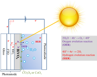
Figure-1: Photoelectrochemical cell and water splitting reactions.
The recombination dynamics of BiVO4 is one of the main obstacle for the water oxidation. Although there are proposed models for the recombination dynamics of bare and doped BiVO4 [1-2], there is a lack of data concerning environmental and operational conditions. Utilizing the Ultrafast transient absorption spectroscopy (UTAS) in operando conditions to understand the charge transfer dynamics shows encouraging results.
In order to have a sufficient water oxidation efficiency, the surface engineering of BiVO4, such as co-catalyst and/or doping, is inevitable. However, the deficiency of a theory or a robust model for the dynamics of intermediate states in operational conditions limits scientists to benefit from the surface modifications. In UTAS, negative absorption difference corresponds to ground state bleach, while the positive features are believed to be hole absorption for some metal-oxide semiconductors. Dynamics of intermediate states can be determined by UTAS. The hole-scavengers are commonly utilized in kinetic measurements and their effect on the dynamics can be noticed in Figure 2. UTAS behavior of BiVO4 forms at later times for the sample in scavenger which indicates the mid-gap states existence.
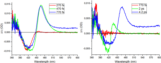
Figure-2: Time-evolution spectra of bare BiVO4 in air (left) and in hole scavenger (Na2SO3)(right).
In this study, we investigate the charge carrier dynamics of CoOx/BiVO4 and N-doped BiVO4 photoanode by utilizing UTAS. The former is believed to affect both catalytic activity of oxidation and suppression of recombination, which is a hot debate [3]. The latter improved the photon absorption and charge transport of BiVO4 [4]. This study aims to gain information of CoOx and nitrogen states on BiVO4 surface in both air and electrolyte via UTAS. Furthermore, we work on these samples in running cell conditions to determine the accurate role of such surface modifications on photo-electrochemical performance for BiVO4 based photoanode. Improved lifetimes of CoOx/BiVO4 and N-doped BiVO4 in different reaction environments will be presented, along with a proposed model for transition dynamics.
References:
1) Ravensbergen et al, J. Phys. Chem. C 2014, 118, 27793−27800
2) Pattengale et al, J. Phys. Chem. C 2016, 120, 1421−1427
3) Zachaus et al, Chem. Sci., 2017, 8, 3712–3719
4) Kim, T. W. et al, Nat. Commun. 6, 8769 (2015)
Coherent diffractive imaging with X-ray lasers opened a bright avenue for determining atomic structure of biomolecules. It uses ultrashort and intense X-ray pulses to take snapshot pictures of biological samples, single particles or protein crystals, before these turn into a plasma due to the extreme radiation dose, aka diffraction before destruction. During destruction the atoms will ionize and emit X-ray fluorescence and this fluorescence can be used to gain further information. In Serial Femtosecond Crystallography (SFX), it can be used to quickly determine successful crystal hits and mitigate a high-rate diffractive data deluge expected at the coming high-repetition X-ray Free-Electron Lasers. X-ray emission may also be used to gain further structural information, through a novel method called Incoherent Diffractive Imaging (IDI) that measure correlation of fluorescent photons from transient metals. Here we study X-ray fluorescence in a plasma environment driven by ultrafast X-ray pulses. We simulate X-ray lasers interacting with biological materials and study the feasibility for hit-finding of protein crystals in SFX experiments, as well as the requirements for a successful IDI experiment.
To date, experiments at x-ray free-electron lasers have generated a very large amount of raw diffraction data. Whilst current pulsed x-ray laser sources measure diffraction patterns at an already very fast rate, with unprecedented development in the instrumentation this data collection rate is projected to rise at least an order of magnitude over the next few years. These large datasets are typically processed and analyzed by many computational routines either in a sequential fashion or in more complicated feedback loops. Furthermore, these routines sometimes employ ad-hoc parameters that are hand tuned by a human operator.
In face of such large datasets, their accompanying processing workflows, and the large parameter space of the algorithms that compose these workflows, it has become practically impossible for peer-validation of single particle reconstructions.
This difficulty in validation is especially acute when imaging unknown structures or unknown conformations where “a true solution” for structural validation is absent. Moreover, the fact that the orientations of individual diffraction patterns are missing and have to be indirectly reconstructed makes it difficult to decide if a pair of patterns are different because they represent the same particle at different orientations or two particles with different structures.
Here we propose novel approach to validate any single particle reconstruction in orientation space, even though the orientations of individual patterns are unknown. Crucially, this approach is independent of the algorithm or dataset used for reconstructions. To do this, we recast the orientation-recovering process into a key-cracking exercise in cryptography: the “correctness” between two cracked keys can be measured by how consistently they decipher a common set of “sentinel messages”.
Using realistic simulations of the single particle pipeline, we demonstrate how to measure an effective uncertainty in the reconstructions in a self-consistent manner. Furthermore, we show that it is possible to separate the negative impact on reconstructions either due to intrinsic noise in diffraction patterns or insufficient number of patterns.
The characterization of the wavefront dynamics is important for many X-ray free-electron laser (XFEL) experiments, in particular for coherent diffractive imaging (CDI), as the reconstructed image is always the product of the incoming wavefront with the object. An accurate understanding of the wavefront is also important to optimize peak power densities to make use of the tightest possible focal spots. Here, we demonstrate that ptychography can be used to reconstruct the full wavefront profile of each individual pulse at the focus of the Atomic, Molecular and Optical (AMO) endstation at the Linac Coherent Light Source (LCLS). Our method uses the mixed-state formalism to retrieve dominant beam components from an ensemble of pulses, which are then used to reconstruct every individual pulse. Among all pulse characterization techniques, our approach works in conditions closest to those in real applications, since it provides the wavefront profile in the focal plane without the need on any other component in the beam path. When integrated as a routine beam diagnostic tool, the described method will benefit the success of XFEL imaging experiments.
The design of the new monochromator beamline FL23 at FLASH, the Free-electron LASer in Hamburg, is made up of 7 optical elements: an elliptical mirror, 2 variable-line-spaced gratings in diffraction order compensation, a vertical slit, a plane mirror and a Kirkpatrick-Baez (KB) focussing system. The monochromotor is designed to work in the soft X-ray range covering a spectral range from 1-20 nm with a spectral resolution of approximately 2000. The two-grating time-delay compensating monochromator (TDCM) will provide a pulse duration in the range of 20 to 100 fs. The beamline FL23 may be used for a variety of experimental techniques such as EUV-spectrometry, to study molecular excitations, magneto-optics Kerr effects, or Extreme Ultraviolet magnetic circular dichroism.
The design process of such a beamline is complex. Simulations are needed to predict the limits on the radii of curvature of the mirrors, the effects of the substrate profile of the gratings and the threshold tolerances of misaligned angles. In order to have a good agreement between the calculations and the experiments, high accuracy simulation tools are required. Raytracing softwares, like SHADOW, are based on the geometry of the optics and the beam. One can introduce parameters in the simulation such as the surface roughness or the reflectivity of the mirrors. However, they neglect some effects like those due to the diffraction. To fully exploit all great features of FLASH2 and to transport parameters from the source, i.e, temporal and spatial coherence length, one can complement raytracing calculations with wavefront propagation. For this purpose the software WaveProgaGator (WPG) was developed at the European XFEL [L.Samoylova et al., J. of Appl. Crystallograpy (2016) 49,1347-1355]. It uses the Synchrotron Radiation Workshop (SRW) library [Chubar, O., and P. Ellaume. Synchrotron Radiation Workshop (SRW) code] and Python binding for wavefront propagation simulations. By taking into account factors like divergence, coherence time or pulse energy of the source, WavePopaGator calculates diffraction effects, interference due to the dimensions of the optics, etc.
Nanoscale hybrid films manifest a set of properties that can beneficial when used in dye lasers, amplifiers, switching devices, solar cells, OLEDs. Studies on the fabrication of sol-gel optical hybrid SiO2 and TiO2 films for photovoltaic devices or planar optical waveguide are continuously caring attention over the years because this materials have numerous applications in the field of microelectronics and optoelectronics. We have studied the characteristics of thin hybrid films which could be useful to tune the quality of mesoscale surface and developed the self-assembly approach based on the sol-gel method to fabricate of high-quality hybrid nanocomposite films using network-forming oxides such as silica or titanium [1-2]. In the frame of given approach the optically transparent, low roughness hybride silicate and titanium films of the nanoscale thickness have been manufactured and have been studied their luminescence property in the broad concentration range [3]. This property of the nanosсale hybrid films was the one of reason to harness them as laser media [4]. The main target of our work was consisted in studying planar waveguide lasing based on the structured hybrid films at its nanoscale thickness. For both films, we observed lasing, which appears as a radiation of the central beam due to the enhanced luminescence (along with the track) and a lasing that occurs due to the distributed grating feedback of the enhancement created by the oncoming waves of the enhanced luminescence. In case of a TiO2 film, it was found that the radiation that emerges from the strip ends sharply with a narrow spectrum under pumping above 0.2 MW/cm2 what is close to the lasing spectrum of the conventional dye laser. The observed narrow directivity in a transverse direction is very surprising because the diffraction should cause this directivity to be much wider. One possible explanation of the narrow directivity is the formation of a thermal lens caused by the transversal temperature gradient. Our estimations show that the thermal lensing may be effective for thin films even for the low intensity lasers. These results show a possibility for a high-quality lasing on the basic mode of a waveguide that forms the central beam.
1. A. Kazakevičius, D. Peckus, L. Valkunas, E.Leonenko, G.Telbiz and V. Gulbinas Journal of Phys.Chem. C, 119 (2015) 19126−19133.
2. G. Telbiz, S. Bugaychuk, E. Leonenko, L. Derzhypolska, V.Gnatovskiy, Nanoscale Research Letters, 10:196 (2015) 1-7.
3. G.Telbiz, E. Leonenko, M. Dvoynenko, Mol. Cryst. Liquid Cryst., 642 (2017) 74-80.
4. E A Tikhonov, V P Yashchuk and G M Telbiz, Laser Phys., 28 (2018) 045801 (7pp)
We present first XUV-pump/XUV-probe transient absorption spectroscopy experiments conducted at the free-electron laser FLASH. Exploiting the partial temporal coherence of the stochastic light fields, this scheme provides access to transient changes in the XUV spectra which are related to dynamics of electronic bound states on a timescale below the average pulse duration. Those transient changes manifest themselves in time-dependent changes in the spectral structure of the FEL-induced dipole response. Experimentally, we split the FEL beam into approximately equal parts with intensities of ~10$^{13}$ W/cm² and average pulse durations of about 50 to 100 fs using the split-and-delay unit at beamline BL2 [1]. Both pulses, denoted by pump and probe are simultaneously detected after transmission through the neon target, and are separately resolved (offset in space) in our grating-based photon spectrometer (E/$\Delta$E ~10$^3$).
Here, we study the time-dependent XUV spectral response of the neon atom and its doubly-charged ion (Ne$^{2+}$) at photon energies close to 50 eV. The pulse-delay ($\tau$) dependent absorbance of the probe pulse is shown in Fig. 1(a) and exhibits resonance lines due to $^3$P–$^3$D 2p–3d spin-orbit multiplet transitions of Ne$^{2+}$ populated in the presence of the FEL pulse via sequential two-photon absorption. A prominent $\Delta\tau=2.2±0.4$ fs “coherence feature” is imprinted on the spectral lines at $\tau=0$, which can be explained by an enhanced coupling of these states due to overlapping temporal intensity peaks of the almost identical pump and probe replica pulses. The essential feature of reduced absorbance is reproduced by employing a non-perturbative few-level model and stochastic fields to drive the transitions (cf. Fig. 1b).
In the near future, a key application will be the precise characterization of asymmetric Fano line shapes in order to study the impact of intense FEL radiation on electron correlation and Fano interference [2].
![(a) XUV-pump/XUV-probe transient absorption spectroscopy on neon with ~50-eV FEL photon energy and pump/probe intensity on the order of 10$^{13}$ W/cm². A “coherence feature” of reduced absorbance is observed at $\tau=0$ with a width of $\Delta\tau=2.2±0.4$ fs. (b) Non-perturbative few-level simulation of the $^3$P–$^3$D 2p–3d transitions of Ne$^{2+}$ which are identified around 49.3 eV and coupled by the partially coherent [3] FEL fields.](https://img1.picload.org/image/dodllcca/fig.png)
[1] M. Wöstmann, et al., The XUV split-and-delay unit at beamline BL2 at FLASH, J. Phys. B 46, 164005 (2013).
[2] U. Fano, Effects of configuration interaction on intensities and phase shifts, Phys. Rev. 124, 1866 (1961).
[3] T. Pfeifer, et al., Partial-coherence method to model experimental free-electron laser pulse statistics, Opt. Lett. 35, 3441 (2010).
Novel types of experiments with both high spatial and temporal resolution have become possible with the intense and short short-wavelength pulses from free-electron lasers. By measuring the diffracted light of a single non-crystallin nanoparticle, fragile objects such as isolated viruses or rotating superfluid helium nanodroplets can be directly visualized. Due to the short pulse duration also ultrafast dynamics like e.g. laser-induced melting or even electronic processes on the nanoscale can be followed by diffractive imaging.
In our experiments we use clusters and nanodroplets as model systems to probe the light-induced processes. For the study of FEL-induced dynamics, two temporally delayed short-wavelength pulses are used, each creating a diffraction image. Therefore, novel methods to separately detect the diffraction images of the same particle created by pump and probe pulse had to be developed. Two recent approaches via non-collinear detection geometries and the use of two-color pulses will be presented and results will be discussed.
The dynamics of liquid water feature a variety of time scales, ranging from extremely fast ballistic-like thermal motion, to slower molecular diffusion and hydrogen-bond rearrangements. Here, will be presented our recent studies[1] using ultrafast coherent X-ray diffraction to investigate the sub-100fs dynamics of water from ambient conditions down to supercooled temperatures. This novel approach utilizes the inherent capability of X-ray speckle visibility spectroscopy at LCLS to measure equilibrium intermolecular dynamics with lengthscale selectivity, by measuring oxygen motion in momentum space.
The observed decay of the speckle contrast at the first diffraction peak, which reflects tetrahedral coordination, is attributed to motion on a molecular scale within the first 120 fs. Through comparison with molecular dynamics simulations, we conclude that the slowing down upon cooling from 328 K down to 253 K is not due to simple thermal ballistic-like motion, but that cage effects play an important role even on timescales over 25 fs due to hydrogen-bonding.
[1] F. Perakis et al., “Coherent X-rays reveal the influence of cage effects on ultrafast water dynamics,” Nat. Commun., vol. 9, no. 1, p. 1917, (2018).
Due to the coherence, high brightness and intensity of laser light sources, the observation and exploitation of non-linear effects in light-matter interaction has been a striving research field in the last decades. Now however, free electron lasers (FELs) at soft and hard x-ray wavelengths are operational which can deliver x-ray fluences on a sample that allow to study non-linear effects in this short wavelength regime as well. Often times though, studies in this field are hampered by the fact that the actual fluence distribution on a sample in a focal spot is not well quantified. This is due to the fact that in non-seeded FELs, there are substantial variations in the pulse intensity and pointing on a shot-to-shot basis. Furthermore, the focal spot typically has non-trivial internal structure due to interference effects, which can change with pointing variations. This calls for a shot-by-shot characterization of the fluence and – ideally – its distribution on the sample. Often, even the integral intensity accepted by a finite sample which is overfilled with the full beam is not easily related to the integral pulse intensity, which is typically available via standard monitors at FEL facilities.
For solid, planar samples studied in transmission, we report on a fluence mapping concept, which allows to monitor the actual x-ray fluence on the sample either in an integral fashion [1] or in a spatially resolved way, i.e. a single shot fluence map can be recorded together with e.g. a diffraction signal from the same illuminated area.[2] The approach is based on fabricating diffraction gratings either directly on the transmissive sample itself or on a separate membrane, which can be independently positioned in the beam. The imaging properties of the diffractive structure can be chosen such that a magnified image of the fluence distribution is obtained on a downstream 2D detector. The fluence imaging concept is single-shot compatible and allows for real time feedback. It is easy to use not only in coincidence with collecting diffraction signals from samples but also as a stand-alone monitoring measurement e.g. for quick alignment of optical elements or to determine the focal position.
[1] M. Schneider, C. M. Günther, C. von Korff Schmising, B. Pfau, S. Eisebitt, Curved gratings as an integrated photon fluence monitor in x-ray transmission scattering experiments. Opt. Ex. 24, 13091 (2016)
[2] M. Schneider, C. M. Günther, B. Pfau, F. Capotondi, M. Manfredda, M. Zangrando, N. Mahne, L. Raimondi, E. Pedersoli, D. Naumenko, S. Eisebitt, In situ single-shot diffractive fluence mapping for X-ray free-electron laser pulses, Nat. Comm. 9, 214 (2018)
For more than 100 years, X-rays have been used in crystallography to determine the structure of crystals and molecules via coherent diffraction methods. With the advent of accelerator-driven free-electron lasers (FEL) new avenues for high-resolution structure determination are presently explored that go far beyond conventional X-ray crystallography [1-3]. Yet, these techniques rely on coherent scattering, where incoherence due to wavefront distortions or incoherent fluorescence emission - often the predominant scattering mechanism - is generally considered as detrimental. Here we show that methods from quantum imaging, i.e., exploiting higher order intensity correlations, can be used to image the full 1D, 2D and 3D arrangement of sources that scatter incoherent X-ray radiation [4-8]. We discuss a number of properties of the new incoherent diffraction imaging method that are conceptually superior to those of conventional coherent X-ray structure determination and point out that current FELs are ideally suited for the implementation of the approach [7]. We present an experimental demonstration in the soft x-ray domain, where higher-order intensity correlations are used to achieve higher fidelities in the image reconstruction and potentially a sub-Abbe resolution [8]. We also discuss recent experiments aiming at full 3D reconstruction of different samples with atomic resolution using hard x-rays.
References
1. H. N. Chapman et al., Nature Phys. 2, 839 (2006).
2. H. N. Chapman et al., Nature 470, 73 (2011).
3. A. Barty, J. Küpper, H. N. Chapman, Annu. Rev. Phys. Chem. 64, 415 (2013), and references therein.
4. C. Thiel, T. Bastin, J. Martin, E. Solano, J. von Zanthier, G. S. Agarwal, Rev. Lett. 99, 133603 (2007).
5. S. Oppel, T. Büttner, P. Kok, J. von Zanthier, Phys. Rev. Lett. 109, 233603 (2012).
6. A. Classen, F. Waldmann, S. Giebel, R. Schneider, D Bhatti, T. Mehringer, J. von Zanthier,
Phys. Rev. Lett. 117, 253601 (2016).
7. A. Classen, K. Ayyer, H. N. Chapman, R. Röhlsberger, J. von Zanthier, Phys. Rev. Lett. 119, 053401 (2017).
8. Schneider et al., Nature Physics 14, 126 (2018); News and Views, Nature Photonics 12, 6 (2018).
On which timescale do the band structure and spin polarization of a ferromagnet change after femtosecond laser excitation and how do they affect the magnetization dynamics? To answer these questions we perform time-, spin-, and angle-resolved photoemission experiments with optical laser pulses and higher-order harmonic radiation.
We have studied ultrafast demagnetization in the local-moment ferromagnets where equilibration of the laser excited state involves more than one timescale, because optical transitions occur in the valence band but the magnetic moment is dominated by the localized 4f electrons.
Following excitation by ultrashort near-infrared pulses, we directly map the transient exchange splitting of the Gd and Tb valence bands near the center of the bulk Brillouin zone. Simultaneously we record the magnetic linear dichroism of the 4f photoemission line. This allows us to compare the magnetization dynamics of 4f core and 5d6s valence electrons in one measurement. To probe the spin polarization, we utilized the unique magnetic properties of the 5d_z^2 surface states. In spin- and time-resolved photoemission with 6.3-eV laser pulses we confirm that the exchange splitting of the Gd and Tb surface state follows that of the 5d6s valence bands. In contrast, the spin polarization of the surface state appears to reflect the magnetization of the 4f core levels.
In non-metallic solids, heat is carried primarily by acoustic phonons. At cryogenic temperatures, the phonon mean free path (MFP) can be large, and ballistic propagation of heat over macroscopic distances has been well documented in the “phonon imaging” studies. At room temperature, the MFP of thermal phonons is small and heat typically propagates via diffusion: once the thermal conductivity is known one can forget about the phonon MFP and rely on the ubiquitous heat equation. In recent years, the interest to the phonon MFP has greatly intensified in the context of micro/nanoscale thermal transport studies stimulated by practical applications such as thermal management of microelectronic devices and designing low thermal conductivity thermoelectric materials. Size effects and non-Fourier transport in practically important materials such as silicon have been observed at room temperature at surprisingly large (~1 $\mu$m) distances, indicating that long-MFP phonons play a much larger role in heat transport than previously thought. On the other hand, coherent acoustic phonons at frequencies above 1 THz, comparable to the frequencies of heat-carrying thermal phonons, can now be generated by ultrashort laser pulses.
Hitherto optical pump-probe experiment has been the workhorse in studying nanoscale thermal transport as well as coherent phonons. However, optical techniques have a number of limitations; in particular, wave vectors of thermal phonons across the Brillouin zone are not directly accessible to optical excitation and probing. The increasing availability of ultrafast EUV and x-ray sources opens many new avenues for studying phonons. In this talk, I will give an overview of some recent research pertaining to the phonon MFP and involving time-resolved measurements with optical, EUV and x-ray pulses. We will discuss studies of non-diffusive heat conduction at small distances using optical and EUV transient grating techniques, including the recent observation of thermal transport near the ballistic limit in diamond. We will also review experiments aimed at measuring the single mode MFP directly: these involved optical pump-probe measurements of sub-THz and THz coherent phonons, as well as diffuse x-ray scattering by “squeezed” thermal phonon populations. While the ultimate goal of getting phonon MFP data across the entire Brillouin zone has not yet been achieved, a number of insights into phonon-phonon, electron-phonon, and phonon-disorder interactions have been obtained. We will conclude by discussing further prospects for studying phonons using short-wavelength radiation.
Free-electron lasers (FELs) are currently the only ultrashort-pulsed photon sources that can provide sufficient photon flux for time-resolved pump-probe solid-state photoemission measurements in the soft and hard x-ray regime. In principle, FELs enable powerful combinations of photoemission techniques, augmented by femtosecond time resolution, in a single experiment. We particularly envision a pump-probe photoemission experiment in which the electronic structure dynamics in energy-momentum space, the chemical dynamics at atomic sites, and the local structural dynamics around atomic sites are simultaneously tracked at the fundamental timescales of electronic and atomic motion by combined time-resolved ARPES, XPS, and XPD. Intriguingly, when hard x-ray FEL radiation is used, the nonequilibrium electron and lattice dynamics can be probed even in the bulk of materials or at buried interfaces. This is especially useful in complex materials or device-like structures where the complete ultrafast photoemission movie provides direct dynamical information on, e.g., the couplings between electronic and structural degrees of freedom or the interfacial carrier dynamics.
Here, we give a brief overview of the current status of time-resolved photoemission at FELs [1-5]. Specifically, we present results obtained from complex materials such as 1T-TaS2, YbInCu4, VO2, and SrTiO3 at the soft x-ray FEL FLASH and the hard x-ray FEL SACLA, respectively. The results demonstrate the practical viability and possible wider impact of FEL-based time-resolved photoemission, but they also illustrate the need for high-repetition-rate FELs such as FLASH and the European XFEL.
This work is supported by the German Federal Ministry of Education and Research (BMBF) through Project Nos. 05K14FKA and 05K16FK2.
[1] S. Hellmann et al., Phys. Rev. Lett. 105, 187401 (2010).
[2] S. Hellmann et al., New J. Phys. 14, 013062 (2012).
[3] L.-P. Oloff et al., New J. Phys. 16, 123045 (2014).
[4] L.-P. Oloff et al., Sci. Rep. 6, 35087 (2016).
[5] L.-P. Oloff et al., in Hard X-ray Photoelectron Spectroscopy (HAXPES) (Springer International Publishing, Switzerland, 2016), pp. 555-568.
Identifying the degrees of freedom that lead to the emergence of superconductivity in iron-based materials remains the subject of active research. Amongst spin-driven scenarios, it has also been suggested that electron-electron correlations enhance the electron-phonon coupling in iron chalcogenides and related pnictides, but direct experimental verification has been lacking.
Measurements of ultrafast lattice dynamics benefit immensely from the advent of x-ray free-electron lasers, providing coherent femtosecond x-ray pulses with unprecedented brilliance. Using the Linac Coherent Light Source at the SLAC National Accelerator Laboratory, we have tracked the light-induced femtosecond coherent lattice motion in FeSe, which originates from a single optical phonon mode. At same time, photoemission spectroscopy allowed us to monitor the corresponding orbital-resolved, coherent change in the electronic band structure [Science 357, 71 (2017)]. Combining these two time-domain experiments into a “coherent lock-in” measurement in the terahertz regime permits quantifying the electron-phonon coupling strength in FeSe purely from experiments and with high precision. Notably, comparison of the experimentally derived electron-phonon deformation potential with theory reveals a strong enhancement of the coupling strength in FeSe owing to correlation effects.
More generally, the coherent lock-in approach establishes an experimental paradigm for precision measurements of fundamental physical quantities by only relying on a linear, coherent response. Thereby, it provides a purely experimental and model-free technique for unbiased tests of emergent phenomena in correlated materials.
I will describe recent experiments using ultrafast electron diffraction and x-ray free electron lasers to probe light-induced structural dynamics in layered transition metal dichalcogenide materials. In particular the focus of this talk will be terahertz-driven atomic-scale responses in WTe2 and related materials. We show, via the measurement of structure factor changes in >200 Bragg reflections, that terahertz light pulses drive interlayer shear phonon excitations with strains >1% that occur along the transition state separating the orthorhombic and monoclinic phases of the material. Theoretical estimates indicate that this is consistent with a photo-doping-driven stabilization of the monoclinic (1T’) phase of the material, a metastable phase not found in equilibrium. A theoretical model also shows that these shear displacements represent a novel ultrafast and energy efficient means to control the topological phase of the material, including both the possibility for inducing a more robust topological structure or as a means to annihilate all Weyl points of opposite chirality.
The results were taken by the SLAC UED collaboration and LCLS Nucleobase collaboration (see author lists of publications below).
In this talk, I will present molecular dynamics obtained by diffraction of relativistic electrons exemplified on the rotational and vibrational modes of small model systems [1,2]. The results have been obtained at a new femtosecond ultrafast electron diffraction source at SLAC. I will point out electron-specific advantages in diffraction of small molecules and discuss future applications on larger molecular systems. In addition, I will present site specific electronic probing of molecular dynamics accomplished by soft x-ray pulses at the oxygen K-edge. We used this scheme to unequivocally observe a molecular intersystem crossing from a ππ state to a nπ state [3]. Combinations of the two techniques for a more complete dynamical description will be discussed.
[1] J. Yang, M. Guehr, T. Vecchione, M. S. Robinson, R. Li, N. Hartmann, X. Shen, R. Coffee, J. Corbett, A. Fry, K. Gaffney, T. Gorkhover, C. Hast, K. Jobe, I. Makasyuk, A. Reid, J. S. Robinson2, S. Vetter, F. Wang, S. Weathersby, C. Yoneda, M. Centurion, X. Wang, Nature Comm. 7, 11232 (2016)
[2] J. Yang, M. Guehr, X. Shen, R. Li, T. Vecchione, R. Coffee, J. Corbett, A. Fry, N. Hartmann, C. Hast, K. Hegazy, K. Jobe, I. Makasyuk, J. Robinson, M. S. Robinson, S. Vetter, S. Weathersby, C. Yoneda, X. Wang, M. Centurion, Phys. Rev. Lett. 115, 173002 (2016)
[3] T. J. A. Wolf, R. H. Myhre, J. P. Cryan, S. Coriani, R. J. Squibb, A. Battistoni, N. Berrah, C. Bostedt, P. Bucksbaum, G. Coslovich, R. Feifel, K. J. Gaffney, J. Grilj, T. J. Martinez, S. Miyabe, S. P. Moeller, M. Mucke, A. Natan, R. Obaid, T. Osipov, O. Plekan, S. Wang, H. Koch and M. Gühr, Nature Communications 8, 29 (2017)
Time-resolved x-ray tools allow measuring electronic and geometric structure changes. X-Ray emission spectroscopy is sensitive to electronic changes, such as oxidation and spin states, while x-ray absorption fine structure tools deliver information about the local geometric structure around the selected absorbing atom. Combining these tools with forward scattering in one single setup allows to extract simultaneous information about the local to rather global structural changes occurring in the reacting system.
We will present case examples, for which pico- and femtosecond x-ray experiments deliver new insight into evolving dynamic processes, including reactive high-valent iron compounds and a class of spin transition systems.
Finally, all these tools can be combined into one single experimental setup, and the Femtosecond X-Ray Experiments (FXE) Instrument at European XFEL will allow just this, while its early operation phase just started in late Summer 2017. We will present the commissioning status of this new instrument at European XFEL [1] together with early results.
[1] Photon Beam Transport and Scientific Instruments at the European XFEL
T. Tschentscher, C. Bressler, J. Grünert, A. Madsen, A. P. Mancuso, M. Meyer,A. Scherz, H. Sinn, U. Zastrau, Appl. Sci. 7, 592 (2017)
Time-resolved x-ray absorption near edge structure (TR-XANES)1 is one of most promising applications of X-ray free electron lasers (XFELs): X-ray absorption measurements combining XFELs and synchronized optical lasers enable one to probe ultrafast changes in the electronic states and the local structure around the absorbing atom during photoreactions.
Here, we report on our recent TR-XANES study of a prototypical copper(I)-phenanthroline complex, [Cu(dmphen)2]+ (dmphen = 2,9-dimethyl-1,10-phenanthroline)2-4, conducted at SPring-8 Angstrom Compact free-electron Laser (SACLA). In the experiment, femtosecond transient XANES changes were measured at three different incident photon energies of 8979.5 eV, 8985.0 eV, and 8986.5 eV. Using the timing diagnostics5,6 at SACLA, the time resolution was improved to ~70 fs by suppressing influences of a timing jitter. At 8979.5 eV (1s -> 3d transition) and 8985.0 eV (1s -> 4p transition), we clearly observed oscillations originating from coherent molecular vibrations, which damp within 1 ps. On the other hand, at 8986.5 eV (1s -> 4p transition), no oscillatory feature was found. These observations umambiguously indicate that the selection of the incident photon energy is critically important to capture a nuclear wave packet in TR-XANES. We will discuss not only the mode assignments of the observed vibrations, but also the selectivity of TR-XAS to molecular vibrations.
[1] T. Katayama, et. al., Femtosecond x-ray absorption spectroscopy with hard x-ray free electron laser, App. Phys. Lett. 103, 131105 (2013).
[2] M. Iwamura, et. al., Real-time observation of the photoinduced structural change of bis(2,9-dimethyl-1,10-phenanthroline)copper(I) by femtosecond fluorescence spectroscopy: A realistic potential curve of the Jahn-Teller distortion, J. Am. Chem. Soc. 129, 5248-5256 (2007).
[3] M. Iwamura, et. al., Coherent nuclear dynamics in ultrafast photoinduced structural change of bis(diimine)copper(I) complex, J. Am. Chem. Soc. 133, 7728-7736 (2011).
[4] G. Capano, et. al., Probing wavepacket dynamics using ultrafast x-ray spectroscopy, J. Phys. B At. Mol. Opt. Phys. 48. 21, (2015).
[5] T. Katayama, et. al., A beam branching method for timing and spectral characterization of hard X-ray free-electron lasers, Struct. Dyn. 3, 034301 (2016).
[6] T. Sato, et. al., Highly efficient arrival timing diagnostics for femtosecond X-ray and optical laser pulses, Appl. Phys. Express 8, 012702 (2015).
Potential energy surfaces play the central role for understanding molecular and chemical dynamics. For chemical reactions on the electronic ground state potential, transition state theory usually provides a strong foundation for understanding chemical reactivity because intra-well equilibration precedes chemical reaction and reactive trajectories proceed along adiabatic trajectories through the transition state with minimal damping. Chemical reactions on electronic excited state potentials differ fundamentally from those on the electronic ground state potential. Reactions can proceed faster than excited state equilibration and reaction dynamics often depend critically on the location of intersections between Born-Oppenheimer potentials where non-adiabatic effects dictate the transition probabilities between distinct electronic state potential energy surfaces. These so-called conical intersections can be seen as the analog of the transition state for electronic excited state chemical reactions and understanding the reaction mechanism of electronic excited states entails identifying the location of these intersections.
Robustly identifying the location of conical intersections that control the relaxation dynamics of electronic excited states has long been a goal and challenge in the chemical sciences. While ultrafast optical methods have made significant contributions to our understanding of electronic excited state reaction dynamics, the detailed interpretation of such measurements proves challenging for the majority of molecular systems. Addressing this challenge requires the development of more direct and differentiated probes of electronic and nuclear dynamics. With the advent of ultrafast x-ray laser sources, powerful x-ray scattering and spectroscopy tools for characterizing steady state nuclear and electronic structure, can now be extended to measure ultrafast electronic and nuclear dynamics.
We have utilized simultaneous K-alpha K-beta X-ray emission spectroscopy (XES) and X-ray diffuse scattering (XDS) to characterize the excited state dynamics in a range of iron-centered molecular systems, from photo-induced spin crossover in the [Fe(2,2’-bipyridine)3]2+ model system, to the details of charge-separation in the photofunctional iron centered N-heterocyclic carbene systems. Combining the sensitivity of XES to the electronic spin moment on the Fe center with the sensitivity of XDS to the dominant changes in intramolecular nuclear structure places significant constraints on the possible trajectories involved in excited state cascade, demonstrating the ability of ultrafast x-ray methods to characterize electronic excited state dynamics of transition metal complexes with unprecedented detail.
Pohang Accelerator Laboratory X-ray Free Electron Laser (PAL-XFEL) facility has finished the commissioning successfully and started user operation from 2016. The NCI experimental hutch, which is one of the hard X-ray experimental stations, has been designed and prepared to perform serial femtosecond crystallography (SFX) experiments. We will present the current status of the sample environments (including sample delivery and dedicated sample chamber), beamline optics and experiment preparation facilities, which are related with SFX.
We present first XUV-pump/XUV-probe transient absorption spectroscopy experiments conducted at the free-electron laser FLASH. Exploiting the partial temporal coherence of the stochastic light fields, this scheme provides access to transient changes in the XUV spectra which are related to dynamics of electronic bound states on a timescale below the average pulse duration. Those transient changes manifest themselves in time-dependent changes in the spectral structure of the FEL-induced dipole response. Experimentally, we split the FEL beam into approximately equal parts with intensities of ~10$^{13}$ W/cm² and average pulse durations of about 50 to 100 fs using the split-and-delay unit at beamline BL2 [1]. Both pulses, denoted by pump and probe are simultaneously detected after transmission through the neon target, and are separately resolved (offset in space) in our grating-based photon spectrometer (E/$\Delta$E ~10$^3$).
Here, we study the time-dependent XUV spectral response of the neon atom and its doubly-charged ion (Ne$^{2+}$) at photon energies close to 50 eV. The pulse-delay ($\tau$) dependent absorbance of the probe pulse is shown in Fig. 1(a) and exhibits resonance lines due to $^3$P–$^3$D 2p–3d spin-orbit multiplet transitions of Ne$^{2+}$ populated in the presence of the FEL pulse via sequential two-photon absorption. A prominent $\Delta\tau=2.2±0.4$ fs “coherence feature” is imprinted on the spectral lines at $\tau=0$, which can be explained by an enhanced coupling of these states due to overlapping temporal intensity peaks of the almost identical pump and probe replica pulses. The essential feature of reduced absorbance is reproduced by employing a non-perturbative few-level model and stochastic fields to drive the transitions (cf. Fig. 1b).
In the near future, a key application will be the precise characterization of asymmetric Fano line shapes in order to study the impact of intense FEL radiation on electron correlation and Fano interference [2].
[1] M. Wöstmann, et al., The XUV split-and-delay unit at beamline BL2 at FLASH, J. Phys. B 46, 164005 (2013).
[2] U. Fano, Effects of configuration interaction on intensities and phase shifts, Phys. Rev. 124, 1866 (1961).
[3] T. Pfeifer, et al., Partial-coherence method to model experimental free-electron laser pulse statistics, Opt. Lett. 35, 3441 (2010).
Myoglobin is a small protein consisting of a single polypeptide chain of 153 amino acid residues and a heme as its active center. It plays a central role in many biological functions based on detection, transport, release and/or binding of molecular ligands such as O2, CO, NO, CN, etc. The unligated high-spin form (deoxyMb) binds the ligand at the Fe of the heme, leading to a change to the planar low-spin ligated form, which is the origin of the respiratory Tense (T) to Relaxed (R) state of the protein. Since, the latter is invariant, it seems ligation causes differences in spin, electronic configuration and geometric structure that determine the role of each ligand.
Nitrosyl-Myoglobin (MbNO), in particular, is not entirely understood despite its biological relevance as it controls various neurophysiological responses. The ultrafast photodissociation of low spin, planar MbNO leads to the high-spin deoxyMb. However, part of the population undergoes recombination on multiple timescales (from sub-ps to 100s ps) and formation of a high-spin domed ligated MbNO is accepted as one of the intermediates on the way back to the planar form. Previous X-ray absorption studies with 70 ps resolution(1) supported the latter hypothesis, but the nature of the earlier time kinetics is unclear. In particular, is the relaxation back to planar a cascade via spin states or is it due to steric hindrances? In order to elucidate these aspects, we combined femtosecond Fe K-edge X-ray absorption spectroscopy (XAS) with X-ray emission spectroscopy (XES) and X-ray diffuse scattering (XDS) at the FXE beamline of the European-XFEL (Hamburg) and at SACLA (Japan). XAS probes the unoccupied density of states (DOS) and the local structure around the Fe atom, while XES probes the occupied DOS and the spin state of the intermediates, XDS allows to unravel structural changes of the protein structure. XAS is showing a faster rise for the electronic changes (main edge) compared to structural changes (post edge) and the evolution of the doming of the Fe over the first few ps. The XES results show a clear signature of the excited quintet state decaying. We will present our results from these measurements and cast them in the context of on-going studies of biosystems at XFELs.
(1) Silatani, M.; Lima, F. A.; Penfold, T. J.; Rittmann, J.; Reinhard, M. E.; Rittmann-Frank, H. M.; Borca, C.; Grolimund, D.; Milne, C. J.; Chergui, M. NO Binding Kinetics in Myoglobin Investigated by Picosecond Fe K-Edge Absorption Spectroscopy. Proc. Natl. Acad. Sci. 2015, 112 (42), 12922–12927.
With the advent of X-ray Free Electron Lasers (XFELs) femtosecond time resolved experiments employing advanced X-ray probe techniques have become routinely possible in a wide variety of scientific domains. A significant number of these experiments concerns the relaxation dynamics following an externally supplied excitation, which is typically realized by an electromagnetic pump pulse originating from a femtosecond IR laser pulse. In general, repetitive pump-probe cycles are required for different delay values to assemble the overall time trace of the relaxation dynamics. In consequence, identical experimental conditions have to be re-established for each pump-probe cycle, a necessity which may be compromised due to practical aspects like the preparation of a sample's initial state, the realization of identical pump parameters or the presence of a temporal jitter between pump and probe pulses. A fundamental restriction concerns the reproducibility of the process itself, which limits the application of pump-probe techniques to the investigation of the reproducible component of ultrafast dynamics.
To overcome this limitation we have conceived a novel experimental approach, which allows continuous probing of a relaxation process with a single X-ray pulse. For this we employ a Fresnel zone plate to stretch an incoming X-ray pulse while introducing at the same time an angular encoding of the arrival time of the X-rays. In recent experiments realized at FLASH we have demonstrated the feasibility of this technique by following with a single X-ray pulse the laser induced demagnetization dynamics [Buzzi17] thanks to transverse magneto-optic Kerr effect.
In order to extend this technique to the pump-probe spectroscopy field, we have performed this single shot x-ray streaking experiment in a transmission geometry at FERMI through a magnetic thin film. Our results show that we are sensitive in one single shot to XMCD signal variation smaller than 1 %. This opens the path for single shot time resolved spectroscopy study. Furthermore, the systematic study of the dynamics of CoDy alloy with different composition and pump intensities shows that the demagnetization time does not depend on the pump fluence but only on the sample composition. This indicates that the ultrafast demagnetization depends only on the sample magnetic properties and not on the temperature rise induced by the pump, at least in the linear demagnetization regime.
Y. Ovcharenko$^{1,9,*}$, A. LaForge$^2$, B. Langbehn$^1$, O. Plekan$^3$, R. Cucini$^3$, P.Finetti$^3$, P.O’Keeffe$^4$, D. Iablonskyi$^5$, N. Toshiyuki$^6$, K. Ueda$^5$, P. Piseri$^7$, M. DiFraia$^3$, R. Richter$^3$, M.Coreno$^{3,4}$, C.Callegari$^3$, K. C. Prince$^{3,8}$, F.Stienkemeier$^2$, T. Möller$^{1,*}$ and M. Mudrich$^{10}$
$^1$Technische Universität Berlin, Germany; $^2$Universität Freiburg, Germany; $^3$Elettra-Sincrotrone Trieste, Trieste, Italy; $^4$ISM-CNR, Rome, Italy; $^5$Tohoku University, Sendai, Japan; $^6$Kyoto University, Kyoto, Japan; $^7$Università degli Studi di Milano, Milano, Italy; $^8$IOM-CNR TASC Laboratory, Trieste, Italy; $^{9}$ European XFEL GmbH, Hamburg, Germany; $^{10}$ Aarhus University, Aarhus, Denmark
$*$ corresponding authors: yevheniy.ovcharenko@xfel.eu, thomas.moeller@physik.tu-berlin.de
The ionization dynamics of He nanodroplets irradiated by intense femtosecond extreme ultraviolet pulses in the range of ${10^{10} - 10^{11}} W/cm^2$ power density and photon energy of $21.4 eV$ has been investigated by photoelectron spectroscopy. The experimental results are interpreted with the help of numerical simulations based on a system of rate equations of various processes such as multi-step ionization, Interatomic Coulombic Decay (ICD) [1,2], secondary inelastic collisions and desorption of electronically excited atoms from He clusters, as well as electronic relaxation processes. In the case of small He clusters (below 1,000 atoms), resonantly excited He-droplet states decay efficiently and fast to low lying $1s2s^3S$, $1s2s^1S$ and $1s2p^1P$ atomic excited states by droplet-induced intra-band and/or inter-band transitions, and then ICD takes place between pairs of electronically excited states, which is then followed by multi-step photoionization. In the case of a few thousand atoms within the He nanodroplet, a pronounced broadening of the photoelectron spectra is observed and the different lines start to interfere due to an increase of the total number of created ICD electrons.
In large He nanodroplets (> 50,000 atoms), inelastic electron scattering starts to play the main role and a cold, dense plasma is formed [2]. Furthermore, when more than two electronically excited atoms are involved the ionization dynamics develops from simple ICD type processes to collective autoionization (CAI) and higher order CAI and/or thermal electron evaporation processes take place. Our results provide an understanding of how the autoionization develops from low power density, characterized by single sharp photolines, to complex ionisation involving many different processes resulting in a cold dense plasma which emits electrons with broad structured distributions.
[1] A. I. Kuleff et al., Phys. Rev. Lett., vol. 105, p. 043004, 2010.
[2] Y. Ovcharenko et al., Phys. Rev. Lett., vol. 112, p. 073401, 2014.
The generation of intense, multicolor fields in the extreme ultraviolet spectral range at Free Electron Lasers (FELs) opens new perspectives for the characterization and control of nonlinear processes in atoms and molecules. These sources can deliver pulses with the high peak intensities enabling the observation of nonlinear processes. These pulses can also be implemented in XUV-pump-XUV-probe experiments by using suitable delay lines. The seeded FEL FERMI (Trieste, Italy) offers the possibility to synthetize multicolor coherent fields, whose amplitudes and relative phases can be independently controlled. The first
experimental demonstration of the coherent control in the XUV spectral region was reported by combining two harmonics with adjustable relative phase and by measuring the photoelectron angular distribution generated by the single and two-photonionisation process [1]. In the temporal domain, the coherent superposition of two or more coherent harmonics determine a temporal beating, whose characteristics can be adjusted by manipulating he relative phases between the harmonics.
I will present novel results about the complete temporal reconstruction of trains of attosecond pulses composed of several harmonics in the XUV and X-ray spectral
range using FELs. The relative phase between the different harmonics can be indipendently controlled giving the possibility to control with attosecond precision the temporal structure of the attosecond pulse train.
[1] K. Prince et al. Nature Photonics 10, 176-179 (2016).
The development of x-ray free-electron lasers (XFELs) opened up a new extreme regime of light-matter interactions and provide unique novel capabilities for studying the structure and dynamics of biological systems, complex materials, and matter under extreme conditions [1,2]. Particularly intriguing is the domain of hard x-ray wavelengths and highest available intensities, since this combination holds potential for imaging applications to reach atomic spatial resolution. Design and interpretation of such experiments to a large extent relies on our quantitative understanding of how individual atoms within an extended polyatomic system respond to multiphoton x-ray absorption, and by how much the distances between the atoms change during the x-ray pulse.
In this talk I will discuss how rare gas atoms and small polyatomic molecules respond to femtosecond hard x-ray pulses at the intensities exceeding 10$^{19}$ W/cm$^{2}$ [3,4]. For atoms, experimental data in the 5.5–8.3 keV range manifest surprisingly structured charge state distributions, which can be accurately described by the newly developed theoretical model [5]. Combined experimental and theoretical analysis demonstrates the importance of resonant and relativistic effects in multiphoton hard x-ray ionization [3]. For molecules containing a single high-Z element, like iodomethane and iodobenzene, it is found that under ultraintense, hard x-rays, the ionization of a molecule is considerably enhanced compared to an individual heavy atom with the same absorption cross section [4], which is qualitatively different from earlier observations in the soft X-ray domain [6,7] or with weaker hard X-rays [8,9]. This enhancement is driven by ultrafast charge transfer within the molecule, which refills the core holes created in the heavy atom, providing further targets for inner-shell ionization and resulting in the emission of more than 50 electrons during the XFEL pulse. For iodomethane, such extreme ionization and fragmentation process can be simulated by the recently developed XMOLECULE package [10], which provides a detailed time-dependent description of molecular dynamics under ultraintense x-ray pulses [4]. Pulse duration and pulse energy dependence of the data reveal further subtleties of the interplay between x-ray absorption, electronic relaxation and nuclear motion, yielding a comprehensive picture of XFEL interactions with small molecules.
[1] Ch. Bostedt et al., Rev. Mod. Phys. 88, 015007 (2016).
[2] E.A. Seddon et al., Rep. Prog. Phys. 80, 115901 (2017).
[3] B. Rudek et al., Nature Comm., under review (2018).
[4] A. Rudenko et al., Nature 545, 129 (2017).
[5] K. Toyota, S.-K. Son & R. Santra, Phys. Rev. A 95, 043412 (2017).
[6] B. Erk et al., Phys. Rev. Lett. 110, 053003 (2013).
[7] B. Erk et al., Science 345, 288 (2014).
[8] K. Motomura et al., J. Phys. Chem. Lett. 6, 2944 (2015).
[9] K. Nagaya et al., Phys. Rev. X 6, 021035 (2016).
[10] Y. Hao et al., Struct. Dyn. 2, 041707 (2015).
The Low Density Matter (LDM) beamline of the Free Electron Laser FERMI in Trieste has been open to Users since December 2012. Members of the atomic-, molecular-, and cluster-science community worldwide have exploited its high-intensity ultrafast pulses for spectroscopy experiments. FERMI is a seeded source, in fact the only one operating in its wavelength range (100 nm–4 nm), and this results in a number of unique features, notably: stable synchronization, precise tunability, and pulse sculpting.
Based on these features an increasing number of experiments have been designed and performed, ranging from nonlinear light-matter interactions to single-shot imaging. I will present the opportunities offered by the LDM beamline, illustrated by recent results of selected experiments, in particular ultrafast chemical dynamics, and control of the relative phase of commensurate wavelengths. Finally I will discuss future developments.
X-ray free electron lasers potentially have the pulse duration and intensity to probe electron motion on the intrinsic timescales found in atoms and molecules, as characterized by the Bohr orbital period of ~150 attoseconds. While sub-orbital period dynamics for deep inner-shell electrons may still out of reach using direct time-domain probes, readily available few femtosecond x-ray pulses are well suited to characterize the relaxation pathways of the highly energetic hole state created by x-ray photoionization, i.e. the competition between the atomically localized Inner-shell decay (Auger emission, fluorescence), intramolecular charge dynamics and Coulomb explosion.
For studies of molecular inner-shell dynamics, we have taken advantage of accelerator-based developments at LCLS [1,2] that engineer two femtosecond x-ray pulses with adjustable duration, wavelength and time delay to probe the first steps following an inner-shell photoelectron ejection event [3,4]. Inner-shell dynamics in two prototypical molecules have been studied using the x-ray pump/x-ray probe methodology: XeF2 via recoil-ion spectroscopy, and, CO via photoelectron spectroscopy, demonstrating both the potential power and limitations of these methods.
For studies of outer-shell electron dynamics, we use the more standard optical pump/x-ray probe configuration. Here the strong-field optical pump generates a distinctive ion signature by impulsively removing an electron from a high-lying orbital to open an isolated x-ray resonance characteristic of the valence hole that can then be used to track its subsequent dynamics [5,6]. We will report on an XFEL-based experiment that addresses the origins of the long-lived electronic coherence in strong-field ionized water previously observed in all-optical experiments that track the hydrated electron [7].
[1] A.A. Lutman et al., Phys. Rev. Lett. 110, 134801 (2013).emphasized text
[2] A.A. Lutman et al., Nat. Photon. 10, 745 (2016).
[3] A. Picón et al., Nat. Commun. 7:11652 (2016).
[4] A. Al Haddad, G. Doumy, C. Bostedt, et al. to be published.
[5] L. Young et al., Phys. Rev. Lett. 97, 083601 (2006).
[6] E. Gouliemakis et al. Nature 466, 739 (2010).
[7] J. Li et al., J. Phys. Chem. Lett. 4, 3698 (2013).
The development of XFELs has opened up opportunities for studying the dynamics of catalysis and biological enzymes. Intense XFEL pulses enable us to apply both X-ray diffraction and X-ray spectroscopic techniques to dilute systems or small protein crystals. By taking advantage of ultra-bright femtosecond X-ray pulses, one can also collect the data under functional conditions of temperature and pressure, in a time-resolved manner, after initiating reactions, and follow the chemical dynamics during catalytic reactions and electron transfer.
We have developed spectroscopy and diffraction techniques necessary to fully utilize the capability of the XFEL X-rays for a wide variety of metalloenzymes, and to study their chemistry under functional conditions. One of such methods is simultaneous data collection for X-ray crystallography and X-ray spectroscopy, to look at the overall structural changes of proteins and the chemical changes at metal catalytic sites. The sample is photochemically or chemically activated at various time delays to capture reaction intermediates with crystallography and spectroscopy.
We have used the above techniques to study photochemical activation of the water oxidation reaction of the Photosystem II (PSII) multi subunit protein complex, in which the Mn4CaO5 cluster catalyzes the reaction. We report the light-induced structure and electronic state changes of the intermediates during the catalysis. The current status of this research and the mechanistic understanding of this metalloenzyme based on the X-ray techniques is presented.
First lasing at the European XFEL was demonstrated in May last year (2017). From these first photons, remarkable progress has been made in delivering beam to the instruments and conducting first user experiments only four months later in September of the same year. I will present an overview of the Single Particles, clusters and Biomolecules and Serial Femtosecond Crystallography (SPB/SFX) instrument, an instrument largely focused on structure determination of biological specimens. I will present highlights from the commissioning of the instrument, as well as show some of the first results from the early user experiments, including successfully determined structures. Finally, an overview of the future development plans for additional capabilities, including the contributions of the SFX user consortium, will be presented.
Time-resolved (TR) crystallography using X-ray free electron lasers (XFELs) is being established and increasingly applied to proteins for visualizing their structural dynamics as "molecular movies". At SACLA, we have applied this technique to study the dynamics of two respiratory enzymes, bovine cytochrome c oxidase (CcO) and fungal NO reductase (P450nor). CcO is the terminal oxidase of cell respiration that catalyzes the reduction of O2 (substrate) to H2O at the heme-cupper site, coupled with proton pumping across the mitochondrial inner membrane. In this study, with the pump-probe scheme, we tracked the structural dynamics of CcO following photo-dissociation of CO (photolabile inhibitor) from the heme, which mimics the reverse process of substrate binding. As a result, we successfully observed gate opening processes in the proton-pump pathway, coupled with CO dissociation in the µs time domain. Next, we turned our focus to observing enzymatic reactions induced by substrate binding. The pump-probe technique with photosensitive caged substrates, which can release substrates by light illumination, may be useful for this purpose. To test this methodology, we performed TR crystallography with a caged substrate, using P450nor as a simple model enzyme. P450nor is an enzyme that catalyzes the reduction of NO (substrate) to N2O at the heme site in the anaerobic respiration in the fungal mitochondrion. To track the NO reduction reaction, we used caged NO that can release NO by UV illumination with a quantum yield of ~1.4. Although the crystal packing affects the reaction rate, we first captured the P450nor-NO complex (initial intermediate) at 20 ms after the caged-NO photolysis. Observation of the subsequent NO reduction reaction of this enzyme is underway.
XFELs with their ultrashort and highly intense pulses open the sub-ps time domain for time-resolved crystallography using small crystals that can be efficiently photolyzed. This is particularly appealing for the study of photosensitive proteins, which contain a light-absorbing chromophore that allows exploitation of light energy as a resource or as a carrier of information initiating intra- or intercellular signaling. The ultra-fast light-induced events comprising double-bond isomerization have been the subject of intense research for decades and have been spectroscopically well characterized. However, direct structural information on the excited state and intermediate structures necessary to understand the underlying mechanisms has been inaccessible until recently. We present recent insight on the initial events in photoisomerization obtained by time-resolved serial femtosecond crystallography experiments in combination with time-resolved spectroscopy and quantum chemical calculations. This comprehensive mechanistic insight is not only important for the fundamental understanding of light-driven processes but has practical impact on future developments of fluorescent proteins for optical nanoscopy or retinal proteins for optogenetics.
Despite being the subject of numerous shock compression studies, the behavior of silicon under dynamic loading is vigorously debated [1-3]. The few studies that combine shock compression and X-ray diffraction have exclusively focused on ``normal'' X-ray geometry whereby X-rays are collected along the shock propagation direction, consequently sampling numerous strain states at once, greatly complicating both phase identification and studies of phase transition kinetics. Here, we present a novel setup performing in situ X-ray diffraction studies perpendicular to the shock propagation direction at the Matter at Extreme Conditions end station at LCLS. Combining the extremely bright microfocussed X-ray beam with a nanosecond drive laser, we unambiguously determine the character of each wave for the first time.
As a further development, we present the simultaneous combination of phase contrast imaging (PCI) techniques with in situ X-ray diffraction perpendicular to the shock compression direction to investigate multiple-wave features in laser-driven germanium. PCI allows one to take femtosecond snapshots of magnified real-space images of shock waves as they progress though matter. X-ray diffraction perpendicular to the shock propagation direction provides the opportunity to isolate and identify different waves and determine the crystal structure unambiguously. We combine these two powerful techniques simultaneously, by using the same Be lens setup to focus the fundamental beam at 8.2 keV to a size of 1.5 mm on target for PCI and the 3rd harmonic at 24.6 keV to a spot size of 2 um on target for diffraction
References
[1] S. J. Turneaure & Y. M. Gupta, “Real-time x-ray diffraction at the impact surface of shocked crystals” APL, 90, 051905 (2007)
[2] N. L. Colburn et al., “Electrical measurements in silicon under shock‐wave compression” JAP, 43, 5007 (1972)
[3] W. H. Gust & E. B. Royce, “Axial Yield Strengths and Two Successive Phase Transition Stresses for Crystalline Silicon” JAP, 42, 1897 (1971)
An accurate knowledge of the properties of iron and iron alloys at high pressures and temperatures is crucial for understanding and modelling planetary interiors. While Earth-size and Super-Earth Exoplanets are being discovered in increasingly large numbers, access to detailed information on liquid properties, melting curves and even solid phases of iron and iron at the pressures and temperatures of their interiors is still strongly limited. In this context, XFEL sources coupled with high-energy lasers afford unique opportunities to measure microscopic structural properties at far extreme conditions. Also the achievable time resolution allows the shock history and phase transition mechanisms to be followed during laser compression, improving our understanding of the high pressure and high strain experiments.
Here we present recent studies devoted to investigate the solid-solid and solid-liquid transition in laser-shocked iron and iron alloys (Fe-Si, Fe-C and Fe-O alloys) using X-ray diffraction, X-ray diffuse scattering and X-ray absorption spectroscopy. Experiment were performed at the MEC end-station of the LCLS facility at SLAC (USA) and at the EH5 end-station of the SACLA facility (Japan). Detection of the diffuse scattering allowed the identification of the first liquid peak position along the Hugoniot, up to 4 Mbar. The time resolution shows ultrafast (between several tens and several hundreds of picoseconds) solid-solid and solid-liquid phase transitions. Future developments at XFEL facilities will enable detailed studies of the solid and liquid structures of iron and iron alloys as well as out-of-Hugoniot studies and will extend our current knowledge to further extreme pressure.
When solid-density matter is irradiated on 100-fsec timescales with a tightly-focussed x-ray FEL the intensities are such that the electrons are heated to temperatures of several hundred eV or more. Core holes induced in the system via photoionisation by the FEL rapidly fill and, depending on the FEL photon energy and intensity, radiation is only emitted during the FEL pulse itself: the photon energies are too great for thermal emission. In this case a time-integrated spectrum can reveal remarkable information about the state of the solid-density plasma on time-scales shorter than that required for an atom to move much further than a lattice spacing: the heating is truly isochoric.[1]
We report here on experiments where such an x-ray FEL was incident on solid targets. The samples emit copious K − α radiation from the various ion stages present during the heating process. However, the radiation for a given stage is only produced strongly when the photon energy of the incident FEL exceeds the K-edge energy of the ion (or is on a resonance), allowing an accurate measurement of the ionisation energy. We find that the ionisation potential is significantly depressed beyond that predicted by simple models used in many atomics-kinetics calculations,[2] but is in good agreement with calculations based on density-functional-theory.[3] Further recent detailed study of the x-ray spectra emitted by such targets, especially when pumping a K-shell resonance, allows us to glean information on the femtosecond collisional ionisation rates by 'clocking' to the Auger rate.[4]
More recent studies have shown that the x-ray laser is capable of isochorically heating very thin solid density targets, just a few hundred atoms across. The solid-density plasmas produced are extremely close to LTE conditions, and the x-ray laser is sufficiently stable and reproducable that the variation of emission with target thickness allows a measure of both the source function and opacity to be gleaned accurately from the data.[5]
We therefore find that the study of x-ray laser heated solid targets is affording a significant increase in our ability to create, control and probe dense plasmas in regions of temperature and density space where, owing to the competition between thermal and coulomb energies, current understanding is limited.
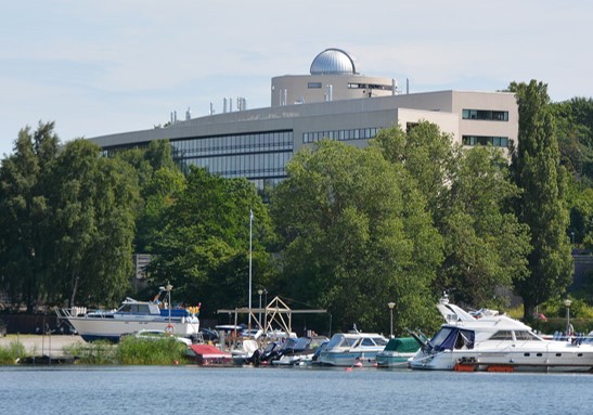 (
(